Quick Summary:

This paper proposes a method to interface Dark-Field X-ray Microscopy (DFXM) with dislocation dynamics modeling by reconstructing three-dimensional deformation gradient tensor fields, enabling non-destructive, in-situ imaging of crystalline deformation. The study validates the approach with simulations, demonstrating accurate reconstruction of dislocation densities and elastic distortion fields, which can guide continuum dislocation dynamics models.

Summary

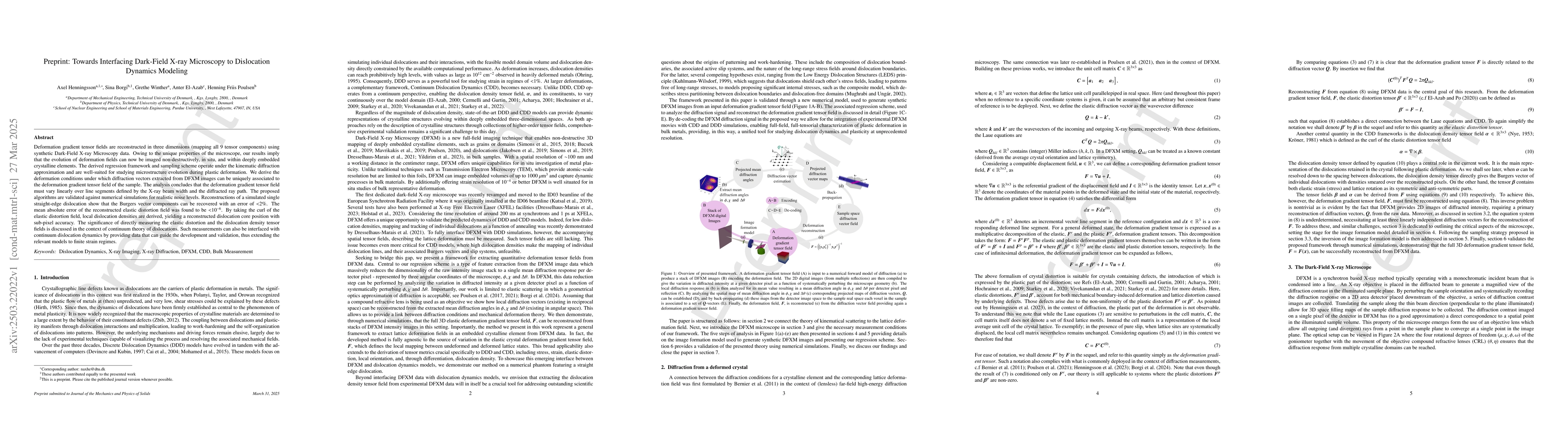
Deformation gradient tensor fields are reconstructed in three dimensions (mapping all 9 tensor components) using synthetic Dark-Field X-ray Microscopy data. Owing to the unique properties of the microscope, our results imply that the evolution of deformation fields can now be imaged non-destructively, in situ, and within deeply embedded crystalline elements. The derived regression framework and sampling scheme operate under the kinematic diffraction approximation and are well-suited for studying microstructure evolution during plastic deformation. We derive the deformation conditions under which diffraction vectors extracted from DFXM images can be uniquely associated to the deformation gradient tensor field of the sample. The analysis concludes that the deformation gradient tensor field must vary linearly over line segments defined by the X-ray beam width and the diffracted ray path. The proposed algorithms are validated against numerical simulations for realistic noise levels. Reconstructions of a simulated single straight-edge dislocation show that the Burgers vector components can be recovered with an error of <2%. The mean absolute error of the reconstructed elastic distortion field was found to be <10^-6. By taking the curl of the elastic distortion field, local dislocation densities are derived, yielding a reconstructed dislocation core position with sub-pixel accuracy. The significance of directly measuring the elastic distortion and the dislocation density tensor fields is discussed in the context of continuum theory of dislocations. Such measurements can also be interfaced with continuum dislocation dynamics by providing data that can guide the development and validation, thus extending the relevant models to finite strain regimes.

AI Key Findings

Generated Jun 10, 2025

Methodology

This research employs synthetic Dark-Field X-ray Microscopy (DFXM) data to reconstruct three-dimensional deformation gradient tensor fields, utilizing a regression framework and sampling scheme under the kinematic diffraction approximation.

Key Results

  • Three-dimensional deformation gradient tensor fields have been reconstructed from synthetic DFXM data, non-destructively, in situ, and within deeply embedded crystalline elements.
  • A regression framework and sampling scheme were derived, validated against numerical simulations, and shown to recover Burgers vector components with an error of <2% and mean absolute error of the reconstructed elastic distortion field of <10^-6.
  • Local dislocation densities were derived by taking the curl of the elastic distortion field, achieving a reconstructed dislocation core position with sub-pixel accuracy.

Significance

This work is significant as it enables the non-destructive imaging of evolving deformation fields in crystalline materials, which can be interfaced with continuum dislocation dynamics models to extend their applicability to finite strain regimes.

Technical Contribution

The paper presents a novel regression framework and sampling scheme for reconstructing three-dimensional deformation gradient tensor fields from synthetic DFXM data, enabling the non-destructive imaging of evolving deformation fields in crystalline materials.

Novelty

The approach combines Dark-Field X-ray Microscopy with dislocation dynamics modeling, offering a new method for studying microstructure evolution during plastic deformation in situ and non-destructively.

Limitations

  • The method is based on the kinematic diffraction approximation, which may not be accurate for large deformations.
  • The reconstruction accuracy depends on the linearity of the deformation gradient tensor field over line segments defined by the X-ray beam width and diffracted ray path.

Future Work

  • Investigate the applicability of the method to larger deformations and more complex microstructures.
  • Explore the integration of real-time DFXM data acquisition and reconstruction for in situ experiments.

Paper Details

PDF Preview

Download PDF

Key Terms

No key terms available for this paper.

Citation Network

Current paper (gray), citations (green), references (blue)

Current Paper
Citations
References
Click to view

No citations found for this paper.

Comments (0)

This website uses cookies to ensure you get the best experience. Learn more