ArxivLens
ArXiv physics.optics

Mid-infrared photothermal imaging flow cytometry

Yusei Sugawara, Keiichiro Toda, Genki Ishigane, Masato Fukushima, Takuro Ideguchi

2606.19842 Published Jun 18, 2026
View PDF

Publication

Published: Jun 18, 2026
Categories: physics.optics

Metrics

Source: ArXiv

Quick Answers

What methodology did the authors use?

Single-shot nanosecond-dual-pulse MIP (SNAP-MIP) microscopy for imaging flow cytometry, encoding MIR-ON and MIR-OFF into separate holographic channels within one camera exposure to achieve ~20 ns temporal separation, enabling wide-field MIP-DH imaging in flowing samples at 500 fps and ~70 events per second. More in Methodology →

What are the key results?

MIP-IFC achieved chemical imaging at 500 frames per second with a cellular event rate of ~70 events s-1. — SNAP-MIP suppression of motion artifacts extended artifact-free flow velocity by five orders of magnitude compared with conventional frame-sequential MIP. More in Key Results →

Why is this work significant?

Presents a label-free, high-throughput chemical imaging platform for single-cell analysis under flow, overcoming motion artifacts and enabling quantitative, multi-parameter phenotyping without fluorescent labels, with potential applications in lipid metabolism, adipogenesis studies, and cellular heterogeneity assessment. More in Significance →

What are the main limitations?

Applicability to all cell types and complex biological media beyond the demonstrated models remains to be validated. — Reliance on specialized SNAP-MIP hardware and synchronization may limit accessibility to broader laboratories. More in Limitations →

Paper Preview

Abstract

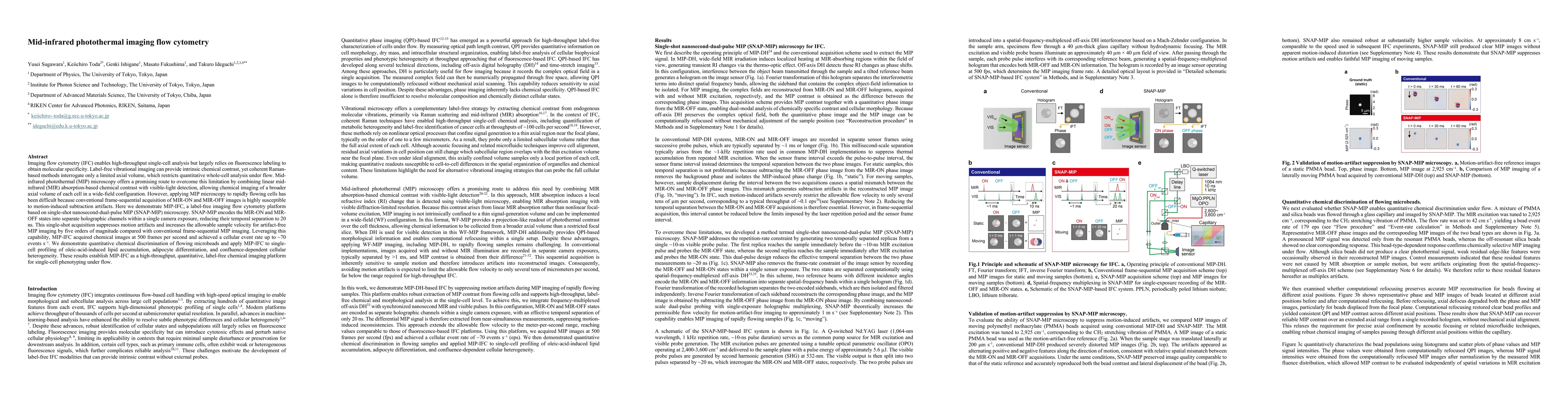
Imaging flow cytometry (IFC) enables high-throughput single-cell analysis but largely relies on fluorescence labeling to obtain molecular specificity. Label-free vibrational imaging can provide intrinsic chemical contrast, yet coherent Raman-based methods interrogate only a limited axial volume, which restricts quantitative whole-cell analysis under flow. Mid-infrared photothermal (MIP) microscopy offers a promising route to overcome this limitation by combining linear mid-infrared (MIR) absorption-based chemical contrast with visible-light detection, allowing chemical imaging of a broader axial volume of each cell in a wide-field configuration. However, applying MIP microscopy to rapidly flowing cells has been difficult because conventional frame-sequential acquisition of MIR-ON and MIR-OFF images is highly susceptible to motion-induced subtraction artifacts. Here we demonstrate MIP-IFC, a label-free imaging flow cytometry platform based on single-shot nanosecond-dual-pulse MIP (SNAP-MIP) microscopy. SNAP-MIP encodes the MIR-ON and MIR-OFF states into separate holographic channels within a single camera exposure, reducing their temporal separation to 20 ns. This single-shot acquisition suppresses motion artifacts and increases the allowable sample velocity for artifact-free MIP imaging by five orders of magnitude compared with conventional frame-sequential MIP imaging. Leveraging this capability, MIP-IFC acquired chemical images at 500 frames per second and achieved a cellular event rate up to ~70 events s^-1. We demonstrate quantitative chemical discrimination of flowing microbeads and apply MIP-IFC to single-cell profiling of oleic-acid-induced lipid accumulation, adipocyte differentiation, and confluence-dependent cellular heterogeneity. These results establish MIP-IFC as a high-throughput, quantitative, label-free chemical imaging platform for single-cell phenotyping under flow.
Key Findings, in focus

Seven facets of this paper, analysed and brought into focus by AI.

AI-generated · verify against source Generated Jun 20, 2026
In focus · one-line synthesis

Presents a label-free, high-throughput chemical imaging platform for single-cell analysis under flow, overcoming motion artifacts and enabling quantitative, multi-parameter phenotyping without fluorescent labels, with potential applications in lipid metabolism, adipogenesis studies, and cellular heterogeneity assessment.

01
MethodologyHow they did it

Single-shot nanosecond-dual-pulse MIP (SNAP-MIP) microscopy for imaging flow cytometry, encoding MIR-ON and MIR-OFF into separate holographic channels within one camera exposure to achieve ~20 ns temporal separation, enabling wide-field MIP-DH imaging in flowing samples at 500 fps and ~70 events per second.

02
ResultsWhat they found
  • MIP-IFC achieved chemical imaging at 500 frames per second with a cellular event rate of ~70 events s-1.
  • SNAP-MIP suppression of motion artifacts extended artifact-free flow velocity by five orders of magnitude compared with conventional frame-sequential MIP.
  • Demonstrated quantitative chemical discrimination in flowing samples and profiling of oleic-acid-induced lipid accumulation, adipocyte differentiation, and confluence-dependent cellular heterogeneity.
  • Integrated MIP contrast with DH-based phase imaging to provide concurrent chemical and morphological information for single-cell phenotyping under flow.
03
SignificanceWhy it matters

Presents a label-free, high-throughput chemical imaging platform for single-cell analysis under flow, overcoming motion artifacts and enabling quantitative, multi-parameter phenotyping without fluorescent labels, with potential applications in lipid metabolism, adipogenesis studies, and cellular heterogeneity assessment.

04
Technical ContributionsWhat's new technically

Development of SNAP-MIP enabling single-shot MIR ON/OFF holographic encoding within one exposure, achieving ~20 ns separation and eliminating motion-induced artifacts in WF-MIP imaging of flowing cells, with demonstrated 500 fps imaging and ~70 eps.

05
NoveltyWhat sets it apart

First demonstration of label-free, high-throughput chemical imaging flow cytometry combining single-shot SNAP-MIP with DH-based detection to probe broad axial volumes in flowing cells, surpassing traditional frame-sequential MIP limitations by several orders of magnitude in allowable flow velocity.

06
LimitationsWhat to be cautious about
  • Applicability to all cell types and complex biological media beyond the demonstrated models remains to be validated.
  • Reliance on specialized SNAP-MIP hardware and synchronization may limit accessibility to broader laboratories.
07
Future WorkWhere it goes next
  • Expand throughput and sampling to diverse primary cell types and clinical samples.
  • Improve spectral specificity to resolve multiple chemical targets simultaneously.
  • Integrate advanced machine learning for automated feature extraction and heterogeneity analysis across large cohorts.
  • Assess compatibility with downstream analyses and multi-omics workflows.

Paper Details

Paper ID: 2606.19842
Categories:
physics.optics

How to Cite This Paper

Reference manager: RIS EndNote

Related Papers

4 found


Discussion 0

This website uses cookies to ensure you get the best experience. Learn more