Publication
Metrics
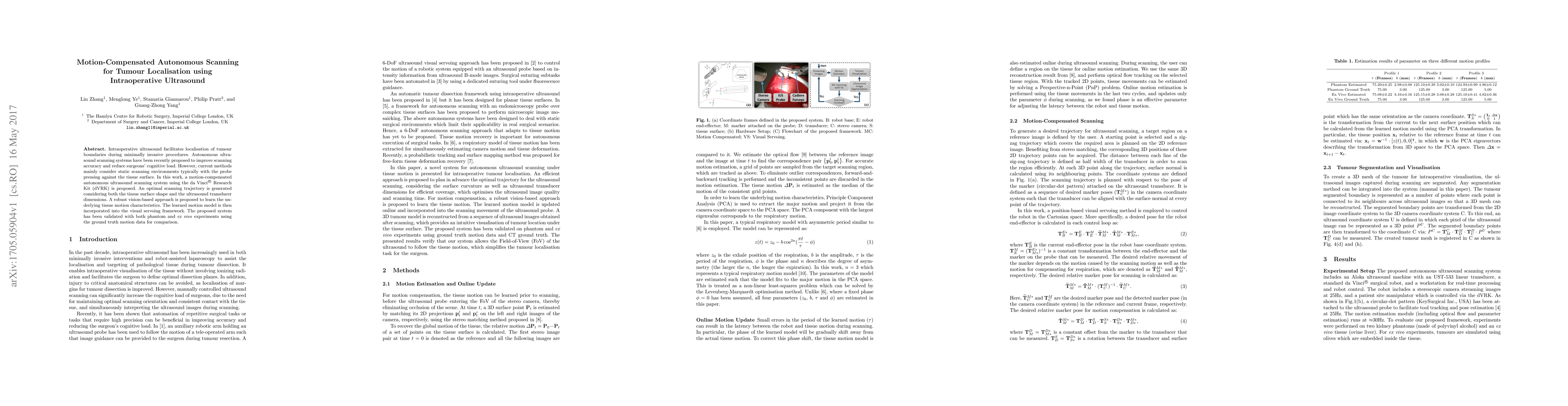
AI Quick Summary
This paper proposes a motion-compensated autonomous scanning system for intraoperative ultrasound to localize tumour boundaries, integrating a vision-based motion model into a visual servoing framework. The system was validated through phantom and ex vivo experiments, demonstrating improved accuracy over static scanning methods.
Paper Preview
Abstract
Intraoperative ultrasound facilitates localisation of tumour boundaries during minimally invasive procedures. Autonomous ultrasound scanning systems have been recently proposed to improve scanning accuracy and reduce surgeons' cognitive load. However, current methods mainly consider static scanning environments typically with the probe pressing against the tissue surface. In this work, a motion-compensated autonomous ultrasound scanning system using the da Vinci Research Kit (dVRK) is proposed. An optimal scanning trajectory is generated considering both the tissue surface shape and the ultrasound transducer dimensions. A robust vision-based approach is proposed to learn the underlying tissue motion characteristics. The learned motion model is then incorporated into the visual servoing framework. The proposed system has been validated with both phantom and ex vivo experiments using the ground truth motion data for comparison.
AI Key Findings
Get AI-generated insights about this paper's methodology, results, significance, and more — seven facets brought into focus.
Impact
Paper Details
PDF Preview
Key Terms
Citation Network
Current paper (gray), citations (green), references (blue)
Display is limited for performance on very large graphs.
Discussion 0