ArxivLens

Quick Summary:

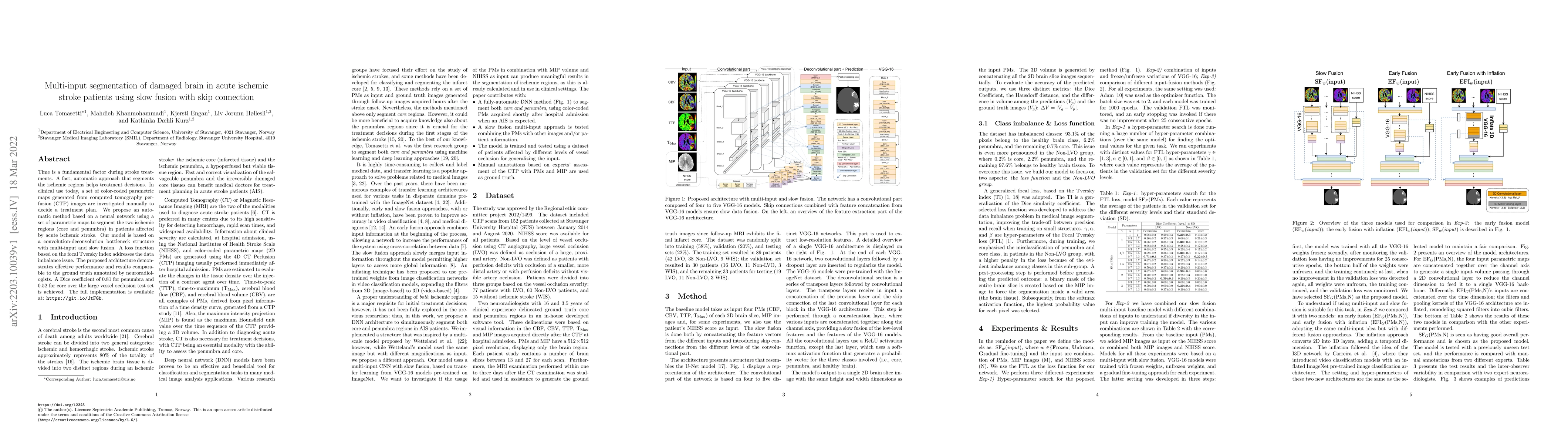
This paper proposes an automatic neural network method for segmenting ischemic regions in acute ischemic stroke patients using multi-input and slow fusion with skip connections. The model achieves a Dice coefficient of 0.81 for penumbra and 0.52 for core, demonstrating effective performance comparable to neuroradiologists.

DOI: 10.7557/18.6223
arXiv Id: 2203.10039
Date Published: 2023-08-22
Date Updated: 2023-08-22

Categories:

Key Terms (TF-IDF):

automatic (0.213)
parametric (0.206)
automatic (0.200)
parametric (0.200)
treatment (0.194)
treatment (0.185)
maps (0.184)
core (0.178)
maps (0.177)
core (0.174)
regions (0.164)
regions (0.162)
set (0.153)
set (0.153)
plan (0.148)
coded (0.147)
plan (0.147)
segment (0.147)
imbalance (0.145)
bottleneck (0.145)
License: http://creativecommons.org/licenses/by/4.0/

Summary:

Time is a fundamental factor during stroke treatments. A fast, automatic approach that segments the ischemic regions helps treatment decisions. In clinical use today, a set of color-coded parametric maps generated from computed tomography perfusion (CTP) images are investigated manually to decide a treatment plan. We propose an automatic method based on a neural network using a set of parametric maps to segment the two ischemic regions (core and penumbra) in patients affected by acute ischemic stroke. Our model is based on a convolution-deconvolution bottleneck structure with multi-input and slow fusion. A loss function based on the focal Tversky index addresses the data imbalance issue. The proposed architecture demonstrates effective performance and results comparable to the ground truth annotated by neuroradiologists. A Dice coefficient of 0.81 for penumbra and 0.52 for core over the large vessel occlusion test set is achieved. The full implementation is available at: https://git.io/JtFGb.

Citation & Reference Network Visualization

This graph shows how this paper is connected to other research. The current paper is shown in dark gray, papers that cite it are in green, and papers it references are in blue. You can:

Current Paper
Papers citing this work (Citations)
Papers cited by this work (References)

• Hover over any node to see the paper title

• Click a node to view that paper's details

• Drag nodes to rearrange the graph

• Use the zoom controls (bottom right) to zoom in/out

Citations and References

Title Authors Year Actions
Vision Transformers for Small Histological Datasets Learned Through Knowledge Distillation
Neel Kanwal, T. Eftestøl, Farbod Khoraminia, T. Zuiverloon, K. Engan
2023
View Paper
CT Perfusion is All we Need: 4D CNN Segmentation of Penumbra and Core in Patients With Suspected Acute Ischemic Stroke
Luca Tomasetti, K. Engan, Liv Jorunn Høllesli, K. D. Kurz, M. Khanmohammadi
2023
View Paper
Self-Supervised Few-Shot Learning for Ischemic Stroke Lesion Segmentation
Luca Tomasetti, Stine Hansen, M. Khanmohammadi, K. Engan, Liv Jorunn Høllesli, K. D. Kurz, Michael C. Kampffmeyer
2023
View Paper
Detection and Localization of Melanoma Skin Cancer in Histopathological Whole Slide Images
Neel Kanwal, Roger Amundsen, Helga Hardardottir, Luca Tomasetti, E. Undersrud, E. Janssen, K. Engan
2023
View Paper
Exploiting 4D CT Perfusion for segmenting infarcted areas in patients with suspected acute ischemic stroke
Luca Tomasetti, K. Engan, Liv Jorunn Høllesli, K. D. Kurz, M. Khanmohammadi
2023
View Paper

Similar Papers

Comments (0)

This website uses cookies to ensure you get the best experience. Learn more