Multimodal post‑mortem analysis of a commercial 26 Ah A123 pouch cell performed in a round‑robin across several laboratories, combining electrochemical tests, flatbed scanner imaging, light and laser scanning microscopy, SEM/EDX, SXES, Raman, FIB‑SEM, X‑ray microscopy, NMR, EPR, GD‑OES, NDP, and ICP‑OES to detect and quantify Li plating.
Multi-Method Li Plating Characterization of a Commercial 26 Ah Li-Ion Pouch-Cell
Publication
Metrics
Quick Answers
What methodology did the authors use?
Multimodal post‑mortem analysis of a commercial 26 Ah A123 pouch cell performed in a round‑robin across several laboratories, combining electrochemical tests, flatbed scanner imaging, light and laser scanning microscopy, SEM/EDX, SXES, Raman, FIB‑SEM, X‑ray microscopy, NMR, EPR, GD‑OES, NDP, and ICP‑OES to detect and quantify Li plating. More in Methodology →
What are the key results?
Li plating appears predominantly at the electrode edges as needle‑shaped dendritic deposits with >70 at % Li confirmed by SEM/EDX and SXES. — Optical methods (scanner, light, laser microscopy) provide rapid, non‑destructive mapping of plating distribution, while spectroscopic techniques confirm metallic Li and distinguish it from intercalated Li. More in Key Results →
Why is this work significant?
The study establishes a validated, multimodal framework for diagnosing Li plating in commercial pouch cells, enabling accurate correlation between electrochemical signatures and physical deposition, which is critical for improving cell safety and lifetime. More in Significance →
What are the main limitations?
Sample preparation and exposure to air can alter surface Li, potentially affecting EDX and spectroscopic results. — Depth‑resolution of optical methods is limited; some spectroscopic techniques (e.g., Raman) suffer from surface scattering and low sensitivity to thin plating layers. More in Limitations →
Abstract
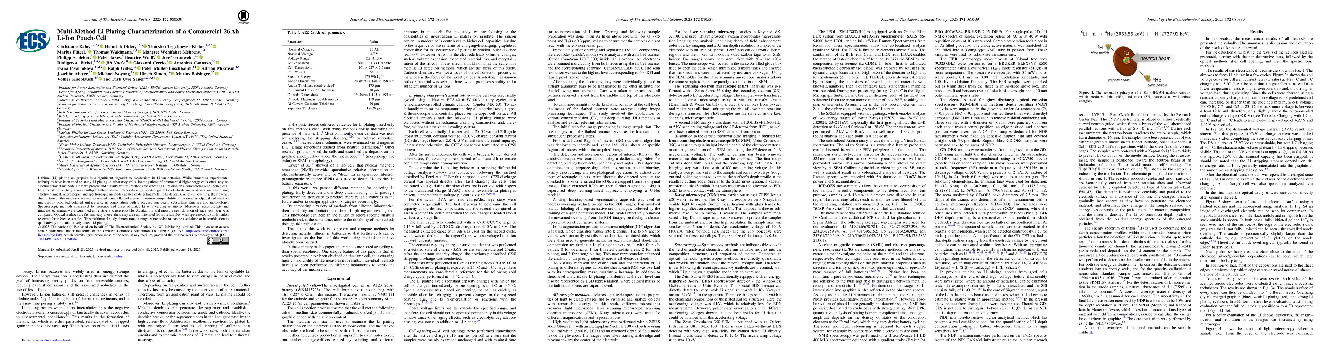
Lithium (Li) plating on graphite is a significant degradation mechanism in Li-ion batteries. While numerous experimental techniques have been used to study Li plating in laboratory cells, investigations of commercial high-energy cells often rely on electrochemical methods. Here we present and classify various methods for detecting Li plating on a commercial A123 pouch cell. In a round robin study across multiple battery research laboratories, Li-plated graphitic electrode material was analyzed using electrochemical, microscopic, and spectroscopic methods capable of detecting metallic Li deposits. After cell opening, their overall distribution on the anode surface was examined using a flatbed scanner to ensure comparability of the samples. Optical and electron microscopy provided detailed surface and, in combination with a focused ion beam, subsurface structure and morphology. Spectroscopic methods confirmed the presence and onset of plated Li with varying sensitivity. Moreover, spectroscopic and imaging techniques were combined correlatively where possible. Availability and measurement duration of each technique was compared. Optical methods are fast and easy to use; thus, they are recommended for most samples, with spectroscopic confirmation reserved for reference samples. This multimodal study demonstrates a range of methods that can be used alone or in combination to qualitatively or quantitatively detect Li-plating.
Key Findings, in focus
Seven facets of this paper, analysed and brought into focus by AI.
The study establishes a validated, multimodal framework for diagnosing Li plating in commercial pouch cells, enabling accurate correlation between electrochemical signatures and physical deposition, which is critical for improving cell safety and lifetime.
- Li plating appears predominantly at the electrode edges as needle‑shaped dendritic deposits with >70 at % Li confirmed by SEM/EDX and SXES.
- Optical methods (scanner, light, laser microscopy) provide rapid, non‑destructive mapping of plating distribution, while spectroscopic techniques confirm metallic Li and distinguish it from intercalated Li.
- Depth profiling (GD‑OES, NDP) shows a surface enrichment of Li (up to 1.6 × 10²² Li atoms cm⁻³) that decreases to bulk levels within 4–8 µm, indicating plating is mainly a surface phenomenon.
The study establishes a validated, multimodal framework for diagnosing Li plating in commercial pouch cells, enabling accurate correlation between electrochemical signatures and physical deposition, which is critical for improving cell safety and lifetime.
A comprehensive, cross‑validated multimodal methodology that correlates surface imaging, elemental analysis, and depth profiling to quantify Li plating in a commercial pouch cell.
First round‑robin multimodal investigation on a commercial 26 Ah pouch cell, combining optical, electron, and spectroscopic techniques with electrochemical data to map plating distribution and depth, providing a benchmark for future diagnostics.
- Sample preparation and exposure to air can alter surface Li, potentially affecting EDX and spectroscopic results.
- Depth‑resolution of optical methods is limited; some spectroscopic techniques (e.g., Raman) suffer from surface scattering and low sensitivity to thin plating layers.
- Integrate rapid optical screening with targeted spectroscopic confirmation to streamline diagnostics in industrial settings.
- Develop in‑situ optical or electrochemical monitoring to capture plating onset in real time and validate simulation models.
Discussion 0