ArxivLens

Quick Summary:

This paper applies deep learning to microscopy to classify colors of single emitters using a standard grayscale camera without additional optical elements, leveraging neural networks to exploit chromatic dependence. The method also demonstrates the use of deep learning to design phase-modulating elements for enhanced color differentiation.

arXiv Id: 1807.01637
Comments: 34 pages, 16 figures
Date Published: 2018-07-05
Date Updated: 2018-07-05

Categories:

Key Terms (TF-IDF):

deep learning (0.248)
deep (0.202)
path (0.199)
elements (0.187)
classification (0.181)
single (0.172)
restoration (0.170)
learning (0.169)
image (0.169)
spectral (0.163)
neural (0.162)
differentiation (0.158)
image classification (0.155)
filters (0.150)
classify (0.145)
molecule (0.145)
colors (0.144)
correctly (0.144)
microscopy (0.138)
camera (0.138)
License: http://arxiv.org/licenses/nonexclusive-distrib/1.0/

Summary:

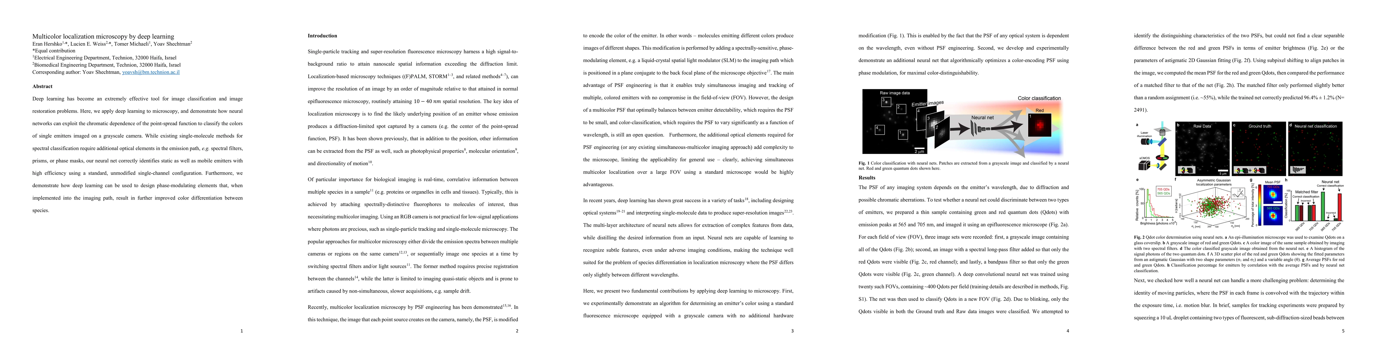
Deep learning has become an extremely effective tool for image classification and image restoration problems. Here, we apply deep learning to microscopy, and demonstrate how neural networks can exploit the chromatic dependence of the point-spread function to classify the colors of single emitters imaged on a grayscale camera. While existing single-molecule methods for spectral classification require additional optical elements in the emission path, e.g. spectral filters, prisms, or phase masks, our neural net correctly identifies static as well as mobile emitters with high efficiency using a standard, unmodified single-channel configuration. Furthermore, we demonstrate how deep learning can be used to design phase-modulating elements that, when implemented into the imaging path, result in further improved color differentiation between species.

Citation & Reference Network Visualization

This graph shows how this paper is connected to other research. The current paper is shown in dark gray, papers that cite it are in green, and papers it references are in blue. You can:

Current Paper
Papers citing this work (Citations)
Papers cited by this work (References)

• Hover over any node to see the paper title

• Click a node to view that paper's details

• Drag nodes to rearrange the graph

• Use the zoom controls (bottom right) to zoom in/out

Citations and References

Title Authors Year Actions
Accelerating multicolor spectroscopic single-molecule localization microscopy using deep learning.
Sunil Kumar Gaire, Yang Zhang, Hongyu Li, Ray Yu, Hao F. Zhang, L. Ying
2020
View Paper
Supercritical Angle Fluorescence Microscopy and Spectroscopy.
M. Oheim, A. Salomon, M. Brunstein
2020
View Paper
Learned sensing: jointly optimized microscope hardware for accurate image classification.
Alex Muthumbi, A. Chaware, Kanghyun Kim, Kevin C Zhou, P. C. Konda, Richard Y. Chen, B. Judkewitz, A. Erdmann, Barbara Kappes, R. Horstmeyer
2019
View Paper
Joint 3D Localization and Classification of Space Debris using a Multispectral Rotating Point Spread Function
Chao Wang, G. Ballard, R. Plemmons, S. Prasad
2019
View Paper
Deep Optics for Monocular Depth Estimation and 3D Object Detection
Julie Chang, Gordon Wetzstein
2019
View Paper
Illumination pattern design with deep learning for single-shot Fourier ptychographic microscopy.
Yi Fei Cheng, Megan Strachan, Zachary Weiss, Moniher Deb, Dawn Carone, V. Ganapati
2018
View Paper
Joint 3 D Localization and Classification of Space Debris using a Multispectral Rotating Point Spread Function
Chao Wang, G. Ballard, R. Plemmons, S. Prasad
2019
View Paper

Comments (0)

This website uses cookies to ensure you get the best experience. Learn more