ArxivLens

Quick Summary:

This paper theoretically derives the single-to-multiple scattering ratio in reflection to predict the penetration depth in optical microscopy, deducing the multiple scattering limit for techniques like confocal microscopy and optical coherence tomography.

Journal Reference: Opt. Exp. 25, 28914-28934, 2017
DOI: 10.1364/OE.25.028914
arXiv Id: 1709.03386
Comments: 18 pages, 7 figures
Date Published: 2017-11-09
Date Updated: 2017-11-09

Key Terms (TF-IDF):

microscopy (0.355)
scattering (0.264)
optical (0.248)
multiple (0.235)
penetration (0.195)
limit (0.189)
tomography (0.182)
quantity (0.177)
quantitatively (0.173)
biological (0.172)
induce (0.168)
reflection (0.168)
coherence (0.165)
insight (0.163)
microscopic (0.161)
offers (0.154)
theoretically (0.150)
depth (0.147)
observation (0.142)
sub (0.141)
License: http://arxiv.org/licenses/nonexclusive-distrib/1.0/

Summary:

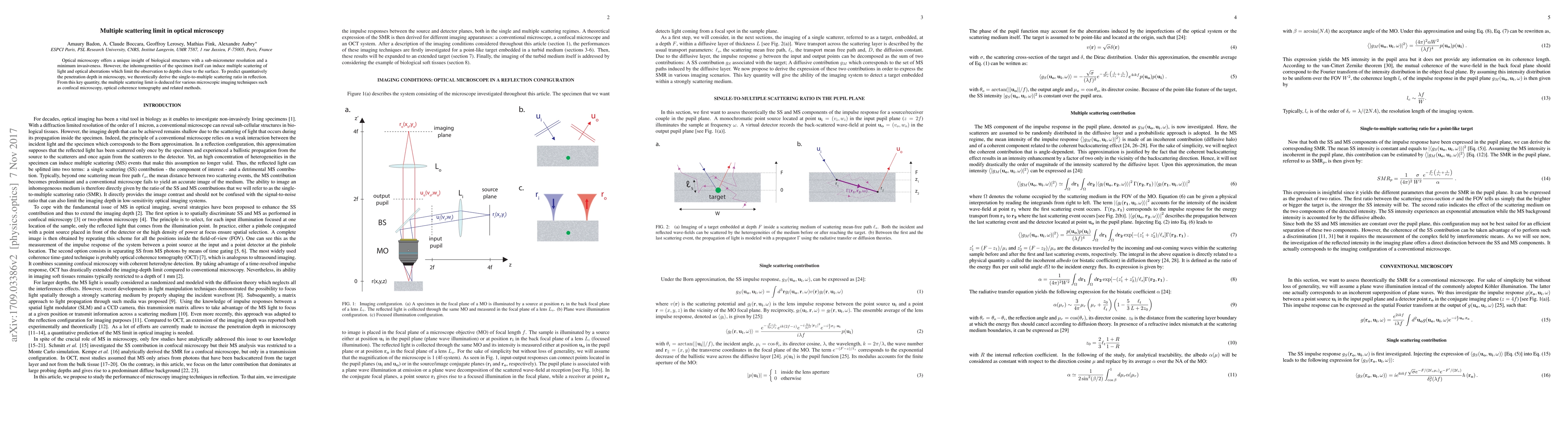
Optical microscopy offers a unique insight of biological structures with a sub-micrometer resolution and a minimum invasiveness. However, the inhomogeneities of the specimen itself can induce multiple scattering of light and optical aberrations which limit the observation to depths close to the surface. To predict quantitatively the penetration depth in microscopy, we theoretically derive the single-to-multiple scattering ratio in reflection. From this key quantity, the multiple scattering limit is deduced for various microscopic imaging techniques such as confocal microscopy, optical coherence tomography and related methods.

Citation & Reference Network Visualization

This graph shows how this paper is connected to other research. The current paper is shown in dark gray, papers that cite it are in green, and papers it references are in blue. You can:

Current Paper
Papers citing this work (Citations)
Papers cited by this work (References)

• Hover over any node to see the paper title

• Click a node to view that paper's details

• Drag nodes to rearrange the graph

• Use the zoom controls (bottom right) to zoom in/out

Citations and References

Title Authors Year Actions
Optical matrix imaging applied to embryology
Victor Barolle, Flavien Bureau, Nicolas Guigui, Paul Balondrade, V. Brochard, Olivier Dubois, A. Jouneau, Am'elie Bonnet-Garnier, Alexandre Aubry
2024
View Paper
Multi-spectral reflection matrix for ultrafast 3D label-free microscopy
Paul Balondrade, Victor Barolle, Nicolas Guigui, Emeric Auriant, Nathan Rougier, C. Boccara, Mathias Fink, A. Aubry
2023
View Paper
Deep imaging inside scattering media through virtual spatiotemporal wavefront shaping
Yiwen Zhang, M. Dinh, Zeyu Wang, Tianhao Zhang, Tianhang Chen, Chia Wei Hsu
2023
View Paper
In-silico clearing approach for deep refractive index tomography by partial reconstruction and wave-backpropagation
Osamu Yasuhiko, Kozo Takeuchi
2023
View Paper
Numerical calculation of near-field in vicinity of three-dimensional nano-optical probes using boundary element method.
Sara Backtiari, M. Sovizi
2022
View Paper
Ultra-parallel label-free optophysiology of neural activity
Rishyashring R. Iyer, Y. Liu, Carlos A. Renteria, Brian E. Tibble, Honggu Choi, Mantas Žurauskas, S. Boppart
2022
View Paper
Nonlinear field-control of terahertz waves in random media for spatiotemporal focusing
V. Cecconi, Vivek Kumar, A. Pasquazi, J. S. Totero Gongora, M. Peccianti
2022
View Paper
Speckle-resolved optical coherence tomography for mesoscopic imaging within scattering media.
M. Cua, Baptiste Blochet, Changhuei Yang
2022
View Paper
Multiple-scattering suppressive refractive index tomography for the label-free quantitative assessment of multicellular spheroids.
Osamu Yasuhiko, Kozo Takeuchi, Hidenao Yamada, Yukio Ueda
2022
View Paper
Deep Penetration Microscopic Imaging with Non-Diffracting Airy Beams
Yong Guo, Yangrui Huang, Jin Li, Luwei Wang, Zhigang Yang, Jinyuan Liu, Xiao Peng, Wei Yan, J. Qu
2021
View Paper
Theory of Multiple Scattering Enhanced Single Particle Plasmonic Sensing
J. Berk, M. Foreman
2021
View Paper
Limiting the incident NA for efficient wavefront shaping through thin anisotropic scattering media
Hyungwon Jin, Byungjae Hwang, Sangwon Lee, Jung‐Hoon Park
2021
View Paper
Three-dimensional substructure imaging of blood cells using maximum entropy tomography based on two non-orthogonal phase images
Hao Han, Ya-Wei Wang, Yang Zou, Jingrong Liao, Y. Xu
2021
View Paper
Optical transfer function of time-gated coherent imaging in the presence of a scattering medium.
Pilsung Kang, Sungsam Kang, Yonghyeon Jo, Hakseok Ko, Guang-Hoon Kim, Ye-Ryoung Lee, W. Choi
2021
View Paper
Deep-tissue label-free quantitative optical tomography
J. van der Horst, A. Trull, J. Kalkman
2020
View Paper
Laser scanning reflection-matrix microscopy for aberration-free imaging through intact mouse skull
Seokchan Yoon, Hojun Lee, J. Hong, Y. Lim, W. Choi
2020
View Paper
Measuring cell displacements in opaque tissues: dynamic light scattering in the multiple scattering regime.
Benjamin Brunel, V. Levy, A. Millet, M. Dolega, A. Delon, R. Pierrat, G. Cappello
2020
View Paper
Deep optical imaging within complex scattering media
Seokchan Yoon, Moonseok Kim, Mooseok Jang, Youngwoon Choi, W. Choi, Sungsam Kang, W. Choi
2020
View Paper
Deep optical imaging within complex scattering media
Seokchan Yoon, Moonseok Kim, Mooseok Jang, Youngwoon Choi, W. Choi, Sungsam Kang, W. Choi
2020
View Paper
Deep tissue space-gated microscopy via acousto-optic interaction
Mooseok Jang, Hakseok Ko, J. Hong, W. Lee, Jae-Seung Lee, W. Choi
2020
View Paper
Distortion matrix approach for ultrasound imaging of random scattering media
William Lambert, L. Cobus, Thomas Frappart, M. Fink, A. Aubry
2019
View Paper
Remote scanning for ultra-large field of view in wide-field microscopy and full-field OCT.
G. Recher, P. Nassoy, A. Badon
2019
View Paper
Computed optical coherence microscopy of mouse brain ex vivo
Meiqi Wu, David M. Small, N. Nishimura, S. Adie
2019
View Paper
Distortion matrix concept for deep optical imaging in scattering media
A. Badon, Victor Barolle, K. Irsch, K. Irsch, A. Boccara, M. Fink, A. Aubry
2019
View Paper
Distortion matrix concept for deep imaging in optical coherence microscopy
A. Badon, Victor Barolle, K. Irsch, A. Boccara, M. Fink, A. Aubry
2019
View Paper
Laser scanning reflection-matrix microscopy for label-free in vivo imaging of a mouse brain through an intact skull.
Seokchan Yoon, Hojun Lee, J. Hong, Y. Lim, W. Choi
2019
View Paper
Modeling of full-field optical coherence tomography in scattering media.
U. Tricoli, R. Carminati
2019
View Paper
Diffraction-limited axial scanning in thick biological tissue with an aberration-correcting adaptive lens
Katrin Philipp, Florian Lemke, S. Scholz, U. Wallrabe, M. Wapler, N. Koukourakis, J. Czarske
2018
View Paper
Coherent space-gated microscopy: a step towards deep-tissue phase imaging of biological cells
Mooseok Jang, Hakseok Ko, W. Lee, Jae-Seung Lee, W. Choi
2018
View Paper
En-face Tomographic Imaging of Scattering Objects Using Single Broadband Light Emitting Diode Based Full-Field Optical Coherence Microscopy
Tulsi Anna, S. Chakraborty, A. Karmenyan, A. Chiou, W. Kuo
2018
View Paper
Multiple Scattering Enhanced Single Particle Sensing
J. Berk, M. Foreman
2021
View Paper
Modeling multiply scattered light in full-field optical coherence tomographic imaging of biological tissues
Yifeng Tang, Wanrong Gao
View Paper

Comments (0)

This website uses cookies to ensure you get the best experience. Learn more