High-resolution 3D refractive index microscopy of multiple-scattering samples from intensity images
Publication
Metrics
AI Quick Summary
This paper introduces a novel 3D refractive index microscopy technique using computational multi-slice beam propagation to reconstruct high-resolution 3D RI distributions of multiple-scattering samples from intensity images, overcoming limitations of traditional interferometric optical diffraction tomography. Experimental results demonstrate its application on biological samples like cells and worms.
Paper Preview
Abstract
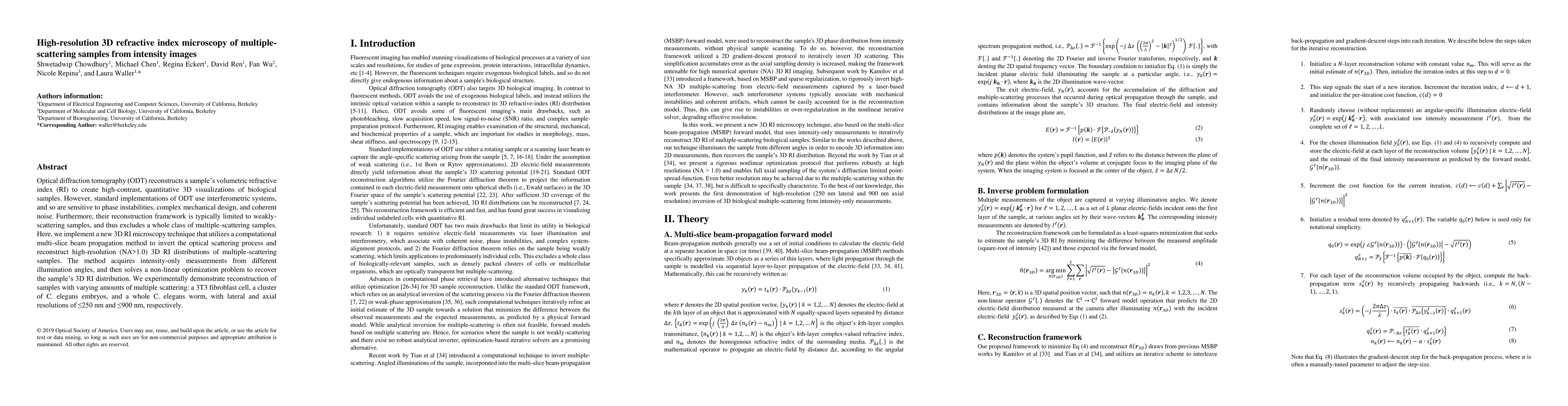
Optical diffraction tomography (ODT) reconstructs a samples volumetric refractive index (RI) to create high-contrast, quantitative 3D visualizations of biological samples. However, standard implementations of ODT use interferometric systems, and so are sensitive to phase instabilities, complex mechanical design, and coherent noise. Furthermore, their reconstruction framework is typically limited to weakly-scattering samples, and thus excludes a whole class of multiple-scattering samples. Here, we implement a new 3D RI microscopy technique that utilizes a computational multi-slice beam propagation method to invert the optical scattering process and reconstruct high-resolution (NA>1.0) 3D RI distributions of multiple-scattering samples. The method acquires intensity-only measurements from different illumination angles, and then solves a non-linear optimization problem to recover the sample 3D RI distribution. We experimentally demonstrate reconstruction of samples with varying amounts of multiple scattering: a 3T3 fibroblast cell, a cluster of C. elegans embryos, and a whole C. elegans worm, with lateral and axial resolutions of 250 nm and 900 nm, respectively.
AI Key Findings
Get AI-generated insights about this paper's methodology, results, significance, and more — seven facets brought into focus.
Impact
Paper Details
PDF Preview
Key Terms
Citation Network
Current paper (gray), citations (green), references (blue)
Display is limited for performance on very large graphs.
Discussion 0