Three-dimensional bi-functional refractive index and fluorescence microscopy (BRIEF)
Publication
Metrics
AI Quick Summary
This paper introduces a novel computational method called BRIEF for simultaneous 3D fluorescence and refractive index (RI) microscopy, enabling label-free RI measurements to correct scattering effects in fluorescence images. The method reconstructs both fluorescence and RI from a single dataset using epi-mode microscopy and demonstrates its effectiveness in improving image quality.
Paper Preview
Abstract
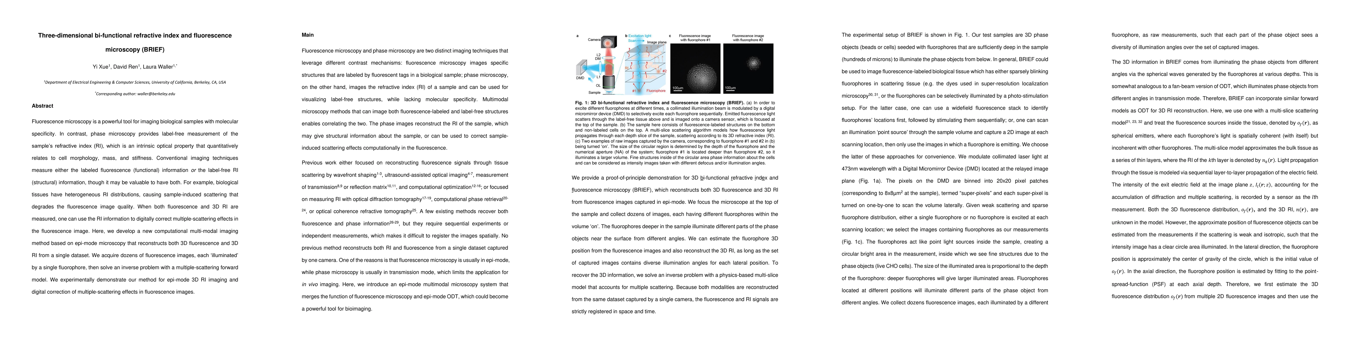
Fluorescence microscopy is a powerful tool for imaging biological samples with molecular specificity. In contrast, phase microscopy provides label-free measurement of the sample's refractive index (RI), which is an intrinsic optical property that quantitatively relates to cell morphology, mass, and stiffness. Conventional imaging techniques measure either the labeled fluorescence (functional) information or the label-free RI (structural) information, though it may be valuable to have both. For example, biological tissues have heterogeneous RI distributions, causing sample-induced scattering that degrades the fluorescence image quality. When both fluorescence and 3D RI are measured, one can use the RI information to digitally correct multiple-scattering effects in the fluorescence image. Here, we develop a new computational multi-modal imaging method based on epi-mode microscopy that reconstructs both 3D fluorescence and 3D RI from a single dataset. We acquire dozens of fluorescence images, each 'illuminated' by a single fluorophore, then solve an inverse problem with a multiple-scattering forward model. We experimentally demonstrate our method for epi-mode 3D RI imaging and digital correction of multiple-scattering effects in fluorescence images.
AI Key Findings
Get AI-generated insights about this paper's methodology, results, significance, and more — seven facets brought into focus.
Impact
Paper Details
Authors
PDF Preview
Key Terms
Citation Network
Current paper (gray), citations (green), references (blue)
Display is limited for performance on very large graphs.
Discussion 0