ArxivLens

Quick Summary:

Structured illumination microscopy (SIM) is shown to achieve dual-modality 3D sub-diffraction resolution for both fluorescence and refractive-index imaging, enabling synergistic exploration of biological mechanisms. The developed SIM framework reconstructs 3D RI/fluorescence visualizations of biological samples, including cancer cells.

arXiv Id: 1708.04693
Comments: 24 pages, 6 figures in main text, 3 figures in supplementary text
Date Published: 2017-08-17
Date Updated: 2017-08-17

Key Terms (TF-IDF):

diffraction (0.285)
biological (0.278)
fluorescence (0.278)
si (0.246)
sub (0.228)
super resolution (0.220)
imaging (0.200)
super (0.175)
resolution (0.174)
coherent (0.173)
experimentally (0.161)
refractive (0.137)
illumination (0.133)
reconstructing (0.131)
modality (0.130)
aperture (0.128)
resolutions (0.128)
demonstration (0.127)
calibration (0.119)
integrate (0.119)
License: http://arxiv.org/licenses/nonexclusive-distrib/1.0/

Summary:

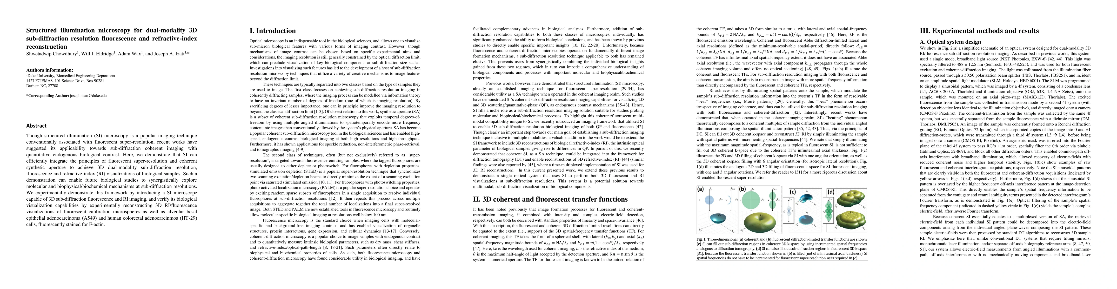
Though structured illumination (SI) microscopy is a popular imaging technique conventionally associated with fluorescent super-resolution, recent works have suggested its applicability towards sub-diffraction coherent imaging with quantitative endogenous biological contrast. Here, we demonstrate that SI can efficiently integrate the principles of fluorescent super-resolution and coherent synthetic aperture to achieve 3D dual-modality sub-diffraction resolution, fluorescence and refractive-index (RI) visualizations of biological samples. Such a demonstration can enable future biological studies to synergistically explore molecular and biophysical/biochemical mechanisms at sub-diffraction resolutions. We experimentally demonstrate this framework by introducing a SI microscope capable of 3D sub-diffraction fluorescence and RI imaging, and verify its biological visualization capabilities by experimentally reconstructing 3D RI/fluorescence visualizations of fluorescent calibration microspheres as well as alveolar basal epithelial adenocarcinoma (A549) and human colorectal adenocarcinmoa (HT-29) cells, fluorescently stained for F-actin.

Citation & Reference Network Visualization

This graph shows how this paper is connected to other research. The current paper is shown in dark gray, papers that cite it are in green, and papers it references are in blue. You can:

Current Paper
Papers citing this work (Citations)
Papers cited by this work (References)

• Hover over any node to see the paper title

• Click a node to view that paper's details

• Drag nodes to rearrange the graph

• Use the zoom controls (bottom right) to zoom in/out

Citations and References

Title Authors Year Actions
Quantitative phase imaging based on holography: trends and new perspectives
Zhengzhong Huang, Liangcai Cao
2024
View Paper
Multiplexing optical diffraction tomography patterns with Fourier synthesis for high-speed volumetric imaging
Peter T. Brown, D. Shepherd
2024
View Paper
Fourier synthesis optical diffraction tomography for kilohertz rate volumetric imaging
Peter T. Brown, Nikta Jabbarzadeh, Aidan Pintuff, Luis Meneses, Ekaterina Monakhova, Rory Kruithoff, N. Wadhwa, Domenico F. Galati, D. Shepherd
2023
View Paper
Single-Shot 3D Topography of Transmissive and Reflective Samples with a Dual-Mode Telecentric-Based Digital Holographic Microscope
A. Doblas, C. Hayes-Rounds, R. Isaac, F. Perez
2022
View Paper
Multicolor structured illumination microscopy and quantitative control of polychromatic light with a digital micromirror device.
Peter T. Brown, Rory Kruithoff, G. Seedorf, D. Shepherd
2021
View Paper
Spatial light interference microscopy: principle and applications to biomedicine.
Xi Chen, M. Kandel, G. Popescu
2020
View Paper
Multicolor structured illumination microscopy and quantitative control of coherent light with a digital micromirror device
Peter T. Brown, Rory Kruithoff, G. Seedorf, D. Shepherd
2020
View Paper
Reduction in data acquisition for resolution improvement in structured illumination digital holographic microscopy
R. Castaneda, T. O’Connor, A. Doblas, B. Javidi
2020
View Paper
Fourier ptychography: current applications and future promises.
P. C. Konda, L. Loetgering, Kevin C Zhou, Shiqi Xu, A. Harvey, R. Horstmeyer
2020
View Paper
High-resolution 3D refractive index microscopy of multiple-scattering samples from intensity images.
Shwetadwip Chowdhury, Michael Chen, Regina Eckert, D. Ren, Fan Wu, N. Repina, L. Waller
2019
View Paper
Speckle-structured illumination for 3D phase and fluorescence computational microscopy.
Li-Hao Yeh, Shwetadwip Chowdhury, N. Repina, L. Waller
2019
View Paper
Resolution enhancement in quantitative phase microscopy
V. Micó, Juanjuan Zheng, Javier García, Z. Zalevsky, P. Gao
2019
View Paper
Deep-learning based three-dimensional label-free tracking and analysis of immunological synapses of chimeric antigen receptor T cells
Moosung Lee, Young-Ho Lee, Jinyeop Song, Geon Kim, YoungJu Jo, YoungJu Jo, Hyun-Seok Min, C. Kim, Y. Park
2019
View Paper
Computational structured illumination for high-content fluorescence and phase microscopy.
Li-Hao Yeh, Shwetadwip Chowdhury, L. Waller
2018
View Paper
Computational multifocal microscopy
K. He, Zihao W. Wang, Xiang Huang, Xiaolei Wang, Seunghwan Yoo, Pablo Ruiz, I. Gdor, A. Selewa, N. Ferrier, N. Scherer, M. Hereld, A. Katsaggelos, O. Cossairt
2018
View Paper
Combining Three-Dimensional Quantitative Phase Imaging and Fluorescence Microscopy for the Study of Cell Pathophysiology
Young Seo Kim, Sangyun Lee, Jaehwang Jung, Seungwoo Shin, HeeSung Choi, Guang-Ho Cha, W. Park, Sumin Lee, Yongkeun Park
2018
View Paper
Low‐coherent optical diffraction tomography by angle‐scanning illumination
KyeoReh Lee, Seungwoo Shin, Z. Yaqoob, P. So, Yongkeun Park
2018
View Paper
Response to Comment on “Is the nuclear refractive index lower than cytoplasm? Validation of phase measurements and implications for light scattering technologies”
Zachary A. Steelman, W. Eldridge, Adam Wax
2018
View Paper
Structured illumination fluorescence microscopy using Talbot self-imaging effect for high-throughput visualization
Shwetadwip Chowdhury, Jeffrey H. Chen, J. Izatt
2018
View Paper
Super-resolution three-dimensional fluorescence and optical diffraction tomography of live cells using structured illumination generated by a digital micromirror device
Seungwoo Shin, Doyeon Kim, Kyoohyun Kim, Yongkeun Park
2018
View Paper
Phase unwrapping by a multi-level grid method for moiré fringes
Yunyun Chen, Chengxing He, Weihao Cheng, Wenzhuo Xie
2025
View Paper
基于边界寻找算法的高温复杂流场区域划分
程伟昊 Cheng Weihao, 陈云云 Chen Yunyun, 陈雅宜 Chen Yayi, 徐梦 Xu Meng
2023
View Paper
Multicolor structured illumination microscopy and quantitative control of polychromatic coherent light with a digital micromirror device
Peter T. Brown, Rory Kruithoff, G. Seedorf, D. Shepherd
2020
View Paper
DeepIS: deep learning framework for three-dimensional label-free tracking of immunological synapses
Moosung Lee, Young-Ho Lee, Jinyeop Song, Geon Kim, YoungJu Jo, Hyun-Seok Min, C. Kim, YongKeun Park
2019
View Paper
Computational fluorescence and phase super-resolution microscopy
Li-Hao Yeh
2019
View Paper

Comments (0)

This website uses cookies to ensure you get the best experience. Learn more