Publication
Metrics
AI Quick Summary
This paper presents a structured illumination microscopy technique that enables multimodal sub-diffraction imaging for both quantitative phase and fluorescence in 3D, significantly enhancing resolution beyond diffraction limits and demonstrating its application in visualizing biological cells.
Paper Preview
Abstract
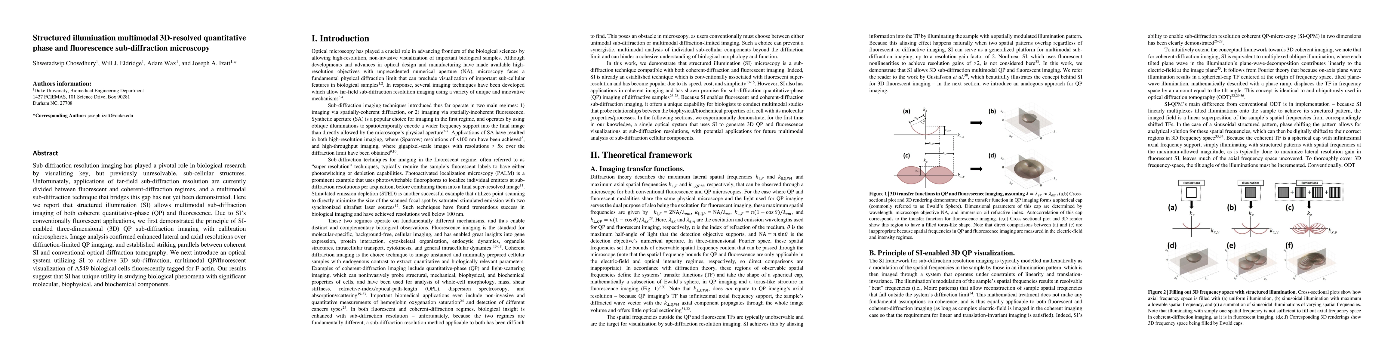
Sub-diffraction resolution imaging has played a pivotal role in biological research by visualizing key, but previously unresolvable, sub-cellular structures. Unfortunately, applications of far-field sub-diffraction resolution are currently divided between fluorescent and coherent-diffraction regimes, and a multimodal sub-diffraction technique that bridges this gap has not yet been demonstrated. Here we report that structured illumination (SI) allows multimodal sub-diffraction imaging of both coherent quantitative-phase (QP) and fluorescence. Due to the conventional fluorescent applications of SI, we first demonstrated the principle of SI-enabled three-dimensional (3D) QP sub-diffraction imaging with calibration microspheres. Image analysis confirmed enhanced lateral and axial resolutions over diffraction-limited QP imaging, and established striking parallels between coherent SI and conventional optical diffraction tomography. We next introduce an optical system utilizing SI to achieve 3D sub-diffraction, multimodal QP/fluorescent visualization of A549 biological cells fluorescently tagged for F-actin. Our results suggest that SI has unique utility in studying biological phenomena with significant molecular, biophysical, and biochemical components.
AI Key Findings
Get AI-generated insights about this paper's methodology, results, significance, and more — seven facets brought into focus.
Impact
Paper Details
PDF Preview
Key Terms
Citation Network
Current paper (gray), citations (green), references (blue)
Display is limited for performance on very large graphs.
Discussion 0