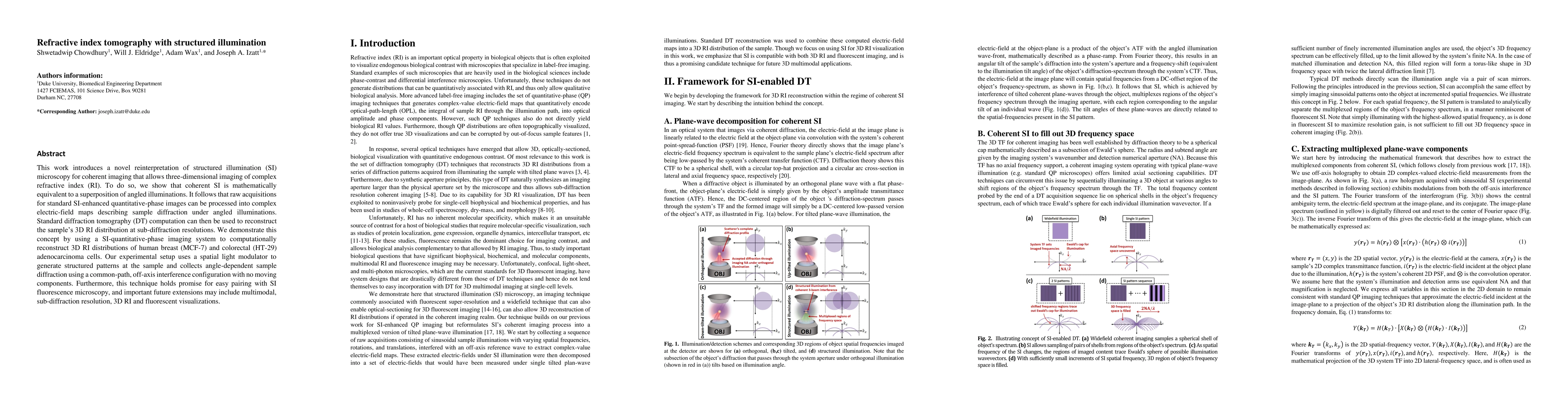
ArxivLens

Quick Summary:

This paper presents a novel method for three-dimensional refractive index tomography using structured illumination microscopy, enabling the reconstruction of complex refractive index distributions at sub-diffraction resolutions. The technique combines structured illumination with diffraction tomography, demonstrating its application on human cancer cells.

DOI: 10.1364/OPTICA.4.000537
arXiv Id: 1702.03595
Comments: 19 total pages (including references), 8 figures
Date Published: 2017-05-11
Date Updated: 2017-05-11

Key Terms (TF-IDF):

diffraction (0.296)
si (0.291)
reconstruct (0.204)
imaging (0.190)
microscopy (0.185)
sample (0.181)
structured (0.173)
coherent (0.164)
quantitative (0.157)
sub (0.154)
refractive (0.131)
fluorescence (0.126)
illumination (0.126)
superposition (0.124)
resolutions (0.122)
processed (0.120)
pairing (0.119)
multimodal (0.119)
complex (0.118)
promise (0.118)
License: http://arxiv.org/licenses/nonexclusive-distrib/1.0/

Summary:

This work introduces a novel reinterpretation of structured illumination (SI) microscopy for coherent imaging that allows three-dimensional imaging of complex refractive index (RI). To do so, we show that coherent SI is mathematically equivalent to a superposition of angled illuminations. It follows that raw acquisitions for standard SI-enhanced quantitative-phase images can be processed into complex electric-field maps describing sample diffraction under angled illuminations. Standard diffraction tomography (DT) computation can then be used to reconstruct the sample 3D RI distribution at sub-diffraction resolutions. We demonstrate this concept by using a SI-quantitative-phase imaging system to computationally reconstruct 3D RI distributions of human breast (MCF-7) and colorectal (HT-29) adenocarcinoma cells. Our experimental setup uses a spatial light modulator to generate structured patterns at the sample and collects angle-dependent sample diffraction using a common-path, off-axis interference configuration with no moving components. Furthermore, this technique holds promise for easy pairing with SI fluorescence microscopy, and important future extensions may include multimodal, sub-diffraction resolution, 3D RI and fluorescent visualizations.

Citation & Reference Network Visualization

This graph shows how this paper is connected to other research. The current paper is shown in dark gray, papers that cite it are in green, and papers it references are in blue. You can:

Current Paper
Papers citing this work (Citations)
Papers cited by this work (References)

• Hover over any node to see the paper title

• Click a node to view that paper's details

• Drag nodes to rearrange the graph

• Use the zoom controls (bottom right) to zoom in/out

Citations and References

Title Authors Year Actions
Structured illumination lensless digital holographic microscopy (SI-LDHM)
Juanjuan Zheng, Xuhong Guo, Ying Ma, Kai Wen, Sha An, Xiaofang Wang, Peng Gao, Jiaming Qian, Chao Zuo
2024
View Paper
DMD and microlens array as a switchable module for illumination angle scanning in optical diffraction tomography
Siqi Yang, Jeongsoo Kim, Mary E Swartz, J. Eberhart, Shwetadwip Chowdhury
2024
View Paper
Quantitative phase imaging based on holography: trends and new perspectives
Zhengzhong Huang, Liangcai Cao
2024
View Paper
Phase Contrast Tomography (PCT)
Ying Ma, Wenjing Feng, Yunze Lei, Lin Ma, Juanjuan Zheng, Sha An, Min Liu, Peng Gao
2024
View Paper
Multiplexing optical diffraction tomography patterns with Fourier synthesis for high-speed volumetric imaging
Peter T. Brown, D. Shepherd
2024
View Paper
Recent Advances and Current Trends in Transmission Tomographic Diffraction Microscopy
N. Verrier, M. Debailleul, O. Haeberlé
2024
View Paper
Fourier synthesis optical diffraction tomography for kilohertz rate volumetric imaging
Peter T. Brown, Nikta Jabbarzadeh, Aidan Pintuff, Luis Meneses, Ekaterina Monakhova, Rory Kruithoff, N. Wadhwa, Domenico F. Galati, D. Shepherd
2023
View Paper
Advances and Overcoming Challenges in Tomographic Diffractive Microscopy
B. Simon, N. Verrier, M. Debailleul, O. Haeberlé
2023
View Paper
Far-field super-resolution chemical microscopy
Mingwei Tang, Yubing Han, Danchen Jia, Qing Yang, Jinmei Cheng
2023
View Paper
Randomness assisted in-line holography with deep learning
Manisha, Aditya Chandra Mandal, Mohit Rathor, Z. Zalevsky, R. Singh
2023
View Paper
Common-path grating-based interferometers with altered temporal and spatial coherence of illumination for quantitative phase microscopy and tomography
M. Trusiak, K. Patorski, P. Zdańkowski, J. Winnik, W. Lipke
2022
View Paper
Multiple-scattering suppressive refractive index tomography for the label-free quantitative assessment of multicellular spheroids.
Osamu Yasuhiko, Kozo Takeuchi, Hidenao Yamada, Yukio Ueda
2022
View Paper
Robust Fourier ptychographic microscopy via a physics-based defocusing strategy for calibrating angle-varied LED illumination.
Chuan-wen Zheng, Shaohui Zhang, Guocheng Zhou, Yao Hu, Q. Hao
2021
View Paper
Resolution Enhancement in Coherent Diffraction Imaging Using High Dynamic Range Image
Yuanyuan Liu, Qingwen Liu, Shuangxiang Zhao, Wenchen Sun, Bingxin Xu, Zuyuan He
2021
View Paper
High-speed multi-view imaging approaching 4pi steradians using conic section mirrors
Kevin C Zhou, Al-Hafeez Z. Dhalla, R. McNabb, Ruobing Qian, Sina Farsiu, J. Izatt
2021
View Paper
Novel approaches to high-speed multi-view imaging approaching 4pi steradians using conic section mirrors: theoretical and practical considerations
Kevin C Zhou, Al-Hafeez Z. Dhalla, R. McNabb, Ruobing Qian, Sina Farsiu, J. Izatt
2021
View Paper
Review of bio-optical imaging systems with a high space-bandwidth product
Jongchan Park, D. Brady, G. Zheng, L. Tian, Liang Gao
2021
View Paper
Common-path intrinsically achromatic optical diffraction tomography.
P. Zdańkowski, J. Winnik, K. Patorski, P. Gocłowski, M. Ziemczonok, M. Józwik, M. Kujawińska, Maciej Trusiak
2021
View Paper
Recent advances in structured illumination microscopy
Ying Ma, Kai Wen, Min Liu, Juanjuan Zheng, K. Chu, Zachary J Smith, Lixin Liu, P. Gao
2021
View Paper
Influence of noise-reduction techniques in sparse-data sample rotation tomographic imaging.
Vinoth Balasubramani, S. Montrésor, H. Tu, Chung-Hsuan Huang, P. Picart, Chau-Jern Cheng
2021
View Paper
Holographic tomography: techniques and biomedical applications [Invited].
Vinoth Balasubramani, A. Kuś, H. Tu, Chau-Jern Cheng, M. Baczewska, W. Krauze, M. Kujawińska
2021
View Paper
Off-axis digital holographic multiplexing for rapid wavefront acquisition and processing
N. Shaked, V. Micó, Maciej Trusiak, A. Kuś, Simcha K. Mirsky
2020
View Paper
Perspectives on liquid biopsy for label‐free detection of “circulating tumor cells” through intelligent lab‐on‐chips
L. Miccio, F. Cimmino, I. Kurelac, M. Villone, V. Bianco, P. Memmolo, F. Merola, M. Mugnano, M. Capasso, A. Iolascon, P. Maffettone, P. Ferraro
2020
View Paper
Cell and nucleus refractive‐index mapping by interferometric phase microscopy and rapid confocal fluorescence microscopy
Shir Cohen Maslaton, I. Barnea, A. Taieb, N. Shaked
2020
View Paper
Multi-layer Born multiple-scattering model for 3D phase microscopy
Michael Chen, D. Ren, Hsiou-Yuan Liu, Shwetadwip Chowdhury, L. Waller
2020
View Paper
Phase-retrieval exact solution based on window modulation
Jianhui Huang, Thomas. T. Gaylord, An Pan, Huiliang Jin, Guoxiang Meng, Q. Ye
2020
View Paper
Reduction in data acquisition for resolution improvement in structured illumination digital holographic microscopy
R. Castaneda, T. O’Connor, A. Doblas, B. Javidi
2020
View Paper
High resolution mapping of subcellular refractive index by Fluorescence Lifetime Imaging: a next frontier in quantitative cell science?
A. Pliss, P. Prasad
2020
View Paper
Computational phase microscopy with modulated illumination
P. Gao, Kai Wen, Lixin Liu, Juanjuan Zheng
2020
View Paper
Digital micromirror device based angle-multiplexed optical diffraction tomography for high throughput 3D imaging of cells
Yanping He, Yijin Wang, R. Zhou
2020
View Paper
Model-based wavefront shaping microscopy.
Abhilash Thendiyammal, Gerwin Osnabrugge, Tom Knop, I. Vellekoop
2020
View Paper
Diffraction tomography with a deep image prior.
Kevin C Zhou, R. Horstmeyer
2019
View Paper
Versatile transmission/reflection tomographic diffractive microscopy approach.
L. Foucault, N. Verrier, M. Debailleul, Jean-Baptiste Courbot, B. Colicchio, B. Simon, L. Vonna, O. Haeberlé
2019
View Paper
High-resolution 3D refractive index microscopy of multiple-scattering samples from intensity images.
Shwetadwip Chowdhury, Michael Chen, Regina Eckert, D. Ren, Fan Wu, N. Repina, L. Waller
2019
View Paper
Optical coherence refraction tomography
Kevin C Zhou, Ruobing Qian, S. Degan, Sina Farsiu, J. Izatt
2019
View Paper
Speckle-structured illumination for 3D phase and fluorescence computational microscopy.
Li-Hao Yeh, Shwetadwip Chowdhury, N. Repina, L. Waller
2019
View Paper
Holographic processing pipeline for tomographic flow cytometry
P. Ferraro, F. Merola, L. Miccio, P. Memmolo, M. Mugnano
2019
View Paper
Resolution enhancement in quantitative phase microscopy
V. Micó, Juanjuan Zheng, Javier García, Z. Zalevsky, P. Gao
2019
View Paper
Computational structured illumination for high-content fluorescence and phase microscopy.
Li-Hao Yeh, Shwetadwip Chowdhury, L. Waller
2018
View Paper
Applications of nanostructures in wide-field, label-free super resolution microscopy
Xiaowei Liu, Chao Meng, Xuechu Xu, Mingwei Tang, Chen-lei Pang, Qing Yang
2018
View Paper
Review on methods of solving the refractive index–thickness coupling problem in digital holographic microscopy of biological cells
Gili Dardikman, N. Shaked
2018
View Paper
Molecular and biophysical analysis of apoptosis using a combined quantitative phase imaging and fluorescence resonance energy transfer microscope
W. Eldridge, Jawad J. Hoballah, A. Wax
2018
View Paper
Low‐coherent optical diffraction tomography by angle‐scanning illumination
KyeoReh Lee, Seungwoo Shin, Z. Yaqoob, P. So, Yongkeun Park
2018
View Paper
Response to Comment on “Is the nuclear refractive index lower than cytoplasm? Validation of phase measurements and implications for light scattering technologies”
Zachary A. Steelman, W. Eldridge, Adam Wax
2018
View Paper
Spectral refractive index assessment of turbid samples by combining spatial frequency near-infrared spectroscopy with Kramers–Kronig analysis
Omri Meitav, Oren Shaul, D. Abookasis
2018
View Paper
Phase contrast tomography at lab on chip scale by digital holography.
F. Merola, P. Memmolo, L. Miccio, M. Mugnano, P. Ferraro
2018
View Paper
Digital holography and its multidimensional imaging applications: a review
T. Tahara, X. Quan, R. Otani, Y. Takaki, O. Matoba
2018
View Paper
Structured illumination fluorescence microscopy using Talbot self-imaging effect for high-throughput visualization
Shwetadwip Chowdhury, Jeffrey H. Chen, J. Izatt
2018
View Paper
Tomographic diffractive microscopy: Towards high-resolution 3-D real-time data acquisition, image reconstruction and display of unlabeled samples
J. Bailleul, B. Simon, M. Debailleul, L. Foucault, N. Verrier, O. Haeberlé
2017
View Paper
Holographic tomography with object rotation and two-directional off-axis illumination.
J. Kostencka, T. Kozacki, M. Józwik
2017
View Paper
Ellipsometry and differential interference contrast microscopic imaging of cellular exoand endocytosis: modelling and experiments
D. Marques
2017
View Paper
Structured illumination microscopy for dual-modality 3D sub-diffraction resolution fluorescence and refractive-index reconstruction.
Shwetadwip Chowdhury, W. Eldridge, A. Wax, J. Izatt
2017
View Paper
Multimodal approach combining optical diffraction tomography and three-dimensional structured illumination microscopy using a digital micromirror device
Seungwoo Shin, Doyeon Kim, Yongkeun Park
2017
View Paper
Reconstructions of refractive index tomograms via a discrete algebraic reconstruction technique.
Moosung Lee, Seungwoo Shin, Yongkeun Park
2017
View Paper
Three‐dimensional correlative single‐cell imaging utilizing fluorescence and refractive index tomography
M. Schürmann, G. Cojoc, S. Girardo, Elke Ulbricht, J. Guck, Paul Müller
2017
View Paper
Tomographic phase microscopy: principles and applications in bioimaging [Invited].
Di Jin, R. Zhou, Z. Yaqoob, P. So
2017
View Paper
Optical diffraction tomography based on quadriwave lateral shearing interferometry
Xun Yuan, Junwei Min, Yuan Zhou, Yuge Xue, Chen Bai, Manman Li, Xiaohao Xu, Baoli Yao
2024
View Paper
Resolution enhancement of digital holographic microscopy via synthetic aperture: a review
P. Gao, Caojin Yuan
2022
View Paper
High-speed multi-view imaging approaching 4pi steradians using conic section mirrors
Evin, Z. C., Hou, H. ’, Afeez, Halla, Uobing, Ian, Ina, Arsiu, oseph, I. Á., Zatt
2021
View Paper
Physics-based correction strategy for large- scale positional deviation of the LED array in Fourier ptychographic microscopy
Huanjian, heng, Haohui, Hang, Uocheng, Hou, Uoan
2021
View Paper
Tomographic Diffractive Microscopy: Principles, Implementations, and Applications in Biology
B. Simon, O. Haeberlé
2019
View Paper
Quantitative Phase Imaging: Principles and Applications
Chenfei Hu, G. Popescu
2019
View Paper
Computational fluorescence and phase super-resolution microscopy
Li-Hao Yeh
2019
View Paper

Comments (0)

This website uses cookies to ensure you get the best experience. Learn more