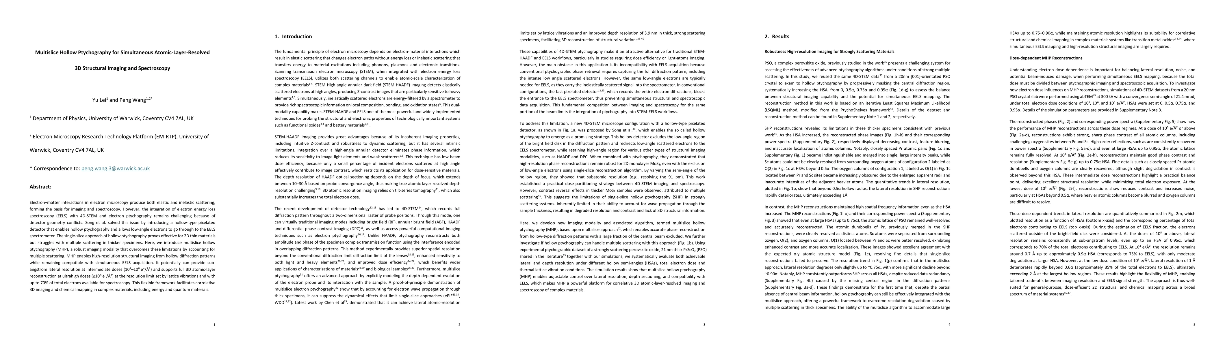
Multislice Hollow Ptychography for Simultaneous Atomic-Layer-Resolved 3D Structural Imaging and Spectroscopy
Publication
Metrics
Paper Preview
Abstract
Electron matter interactions in electron microscopy produce both elastic and inelastic scattering, forming the basis for imaging and spectroscopy. However, the integration of electron energy loss spectroscopy (EELS) with 4D-STEM and electron ptychography remains challenging because of detector geometry conflicts. Song et al. solved this issue by introducing a hollow type pixelated detector that enables hollow ptychography and allows low angle electrons to go through to the EELS spectrometer. The single-slice approach of hollow ptychography proves effective for 2D thin materials but struggles with multiple scattering in thicker specimens. Here, we introduce multislice hollow ptychography (MHP), a robust imaging modality that overcomes these limitations by accounting for multiple scattering. MHP enables high-resolution structural imaging from hollow diffraction patterns while remaining compatible with simultaneous EELS acquisition. It potentially can provide sub-angstrom lateral resolution at intermediate doses and supports full 3D atomic-layer reconstruction at ultrahigh doses, with up to 70% of total electrons available for spectroscopy. This flexible framework facilitates correlative 3D imaging and chemical mapping in complex materials, including interfaces, defects, and dopants.
AI Key Findings
Get AI-generated insights about this paper's methodology, results, significance, and more — seven facets brought into focus.
Impact
Paper Details
Authors
PDF Preview
Citation Network
Current paper (gray), citations (green), references (blue)
Display is limited for performance on very large graphs.
Discussion 0