Publication
Metrics
AI Quick Summary
This paper explores the use of Helium Ion Microscopy (HIM) to create nanopores down to 1.3 nm in materials like silicon nitride, carbon nanomembranes, and graphene. The study employs HIM and high-resolution scanning transmission electron microscopy to characterize and analyze the nanopores, providing insights into the helium ion beam's profile.
Paper Preview
Abstract
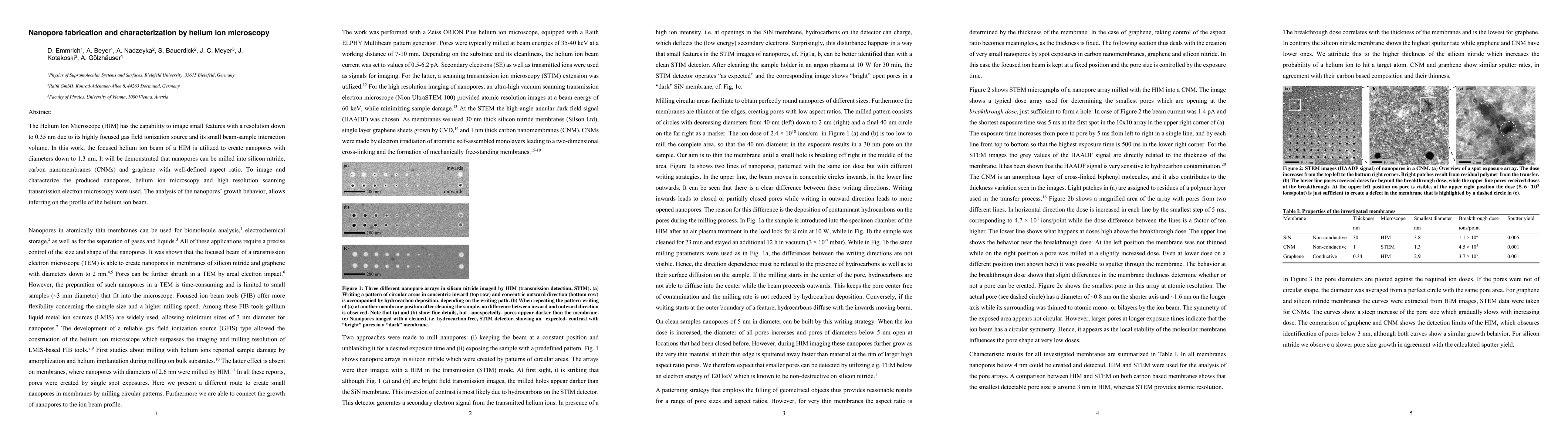
The Helium Ion Microscope (HIM) has the capability to image small features with a resolution down to 0.35 nm due to its highly focused gas field ionization source and its small beam-sample interaction volume. In this work, the focused helium ion beam of a HIM is utilized to create nanopores with diameters down to 1.3 nm. It will be demonstrated that nanopores can be milled into silicon nitride, carbon nanomembranes (CNMs) and graphene with well-defined aspect ratio. To image and characterize the produced nanopores, helium ion microscopy and high resolution scanning transmission electron microscopy were used. The analysis of the nanopore's growth behavior, allows inferring on the profile of the helium ion beam.
AI Key Findings
Get AI-generated insights about this paper's methodology, results, significance, and more — seven facets brought into focus.
Impact
Paper Details
PDF Preview
Key Terms
Citation Network
Current paper (gray), citations (green), references (blue)
Display is limited for performance on very large graphs.
Discussion 0