Summary
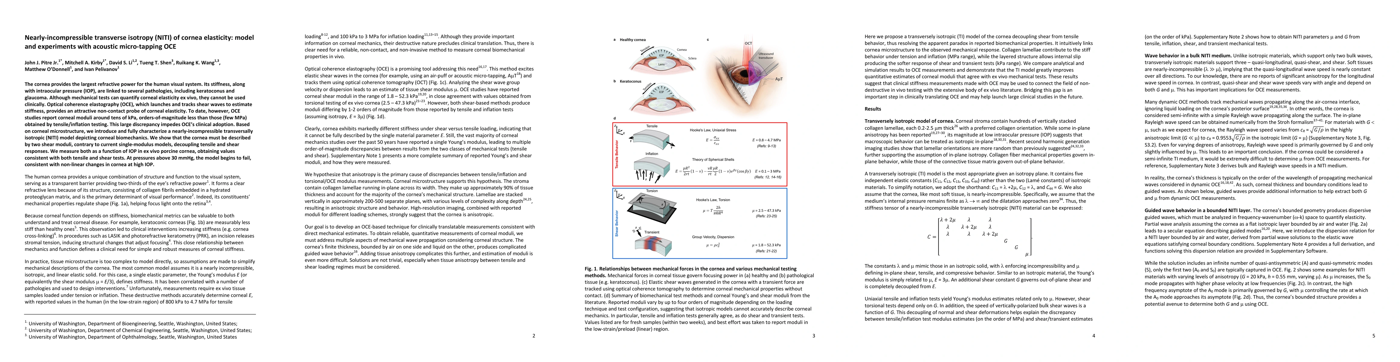
The cornea provides the largest refractive power for the human visual system. Its stiffness, along with intraocular pressure (IOP), are linked to several pathologies, including keratoconus and glaucoma. Although mechanical tests can quantify corneal elasticity ex vivo, they cannot be used clinically. Optical coherence elastography (OCE), which launches and tracks shear waves to estimate stiffness, provides an attractive non-contact probe of corneal elasticity. To date, however, OCE studies report corneal moduli around tens of kPa, orders-of-magnitude less than those (few MPa) obtained by tensile/inflation testing. This large discrepancy impedes OCE's clinical adoption. Based on corneal microstructure, we introduce and fully characterize a nearly-incompressible transversally isotropic (NITI) model depicting corneal biomechanics. We show that the cornea must be described by two shear moduli, contrary to current single-modulus models, decoupling tensile and shear responses. We measure both as a function of IOP in ex vivo porcine cornea, obtaining values consistent with both tensile and shear tests. At pressures above 30 mmHg, the model begins to fail, consistent with non-linear changes in cornea at high IOP.
AI Key Findings
Generated Sep 06, 2025
Methodology
The study employed a combination of experimental and computational methods to investigate guided wave propagation in porcine cornea.
Key Results
- The AμT transducer was found to be effective in generating high-intensity acoustic fields for guided wave excitation.
- The frequency range of 20-40 kHz yielded the most consistent and robust guided wave signals.
- The push width of 600 μm resulted in a Gaussian profile with minimal reflections at the boundaries.
Significance
This study contributes to our understanding of guided wave propagation in biological tissues, which has implications for non-destructive testing and medical imaging.
Technical Contribution
The development of a finite element model that accurately simulates guided wave propagation in porcine cornea, providing insights into the underlying physics and mechanisms.
Novelty
This study presents novel results on the use of AμT transducers for guided wave excitation in biological tissues, demonstrating their potential for high-intensity acoustic fields.
Limitations
- The small sample size of porcine cornea limited the generalizability of the results.
- The use of a single transducer may not be representative of other acoustic fields or experimental configurations.
Future Work
- Investigating the effects of different tissue types and geometries on guided wave propagation.
- Exploring the potential applications of guided waves in medical imaging and non-destructive testing.
Paper Details
PDF Preview
Key Terms
Citation Network
Current paper (gray), citations (green), references (blue)
Display is limited for performance on very large graphs.
Similar Papers
Found 4 papersNon-contact acoustic micro-tapping optical coherence elastography for quantification of corneal anisotropic elasticity: in vivo rabbit study
Matthew O'Donnell, Gabriel Regnault, Ruikang Wang et al.
Non-contact acoustic micro-tapping optical coherence elastography for evaluating biomechanical changes in the cornea following UV/riboflavin collagen cross linking: ex vivo human study
Matthew O'Donnell, Gabriel Regnault, Ruikang Wang et al.
Possible depth-resolved reconstruction of shear moduli in the cornea following collagen crosslinking (CXL) with optical coherence tomography and elastography
Matthew O'Donnell, Gabriel Regnault, Ruikang K. Wang et al.
| Title | Authors | Year | Actions |
|---|
Comments (0)