Publication
Metrics
AI Quick Summary
This study demonstrates the use of non-contact Acoustic micro-Tapping Optical Coherence Elastography (AuT-OCE) to accurately measure corneal elastic moduli in vivo in rabbits, showing strong corneal anisotropy. The method shows promise for clinical translation to improve diagnosis and treatment of ectatic corneal diseases.
Paper Preview
Abstract
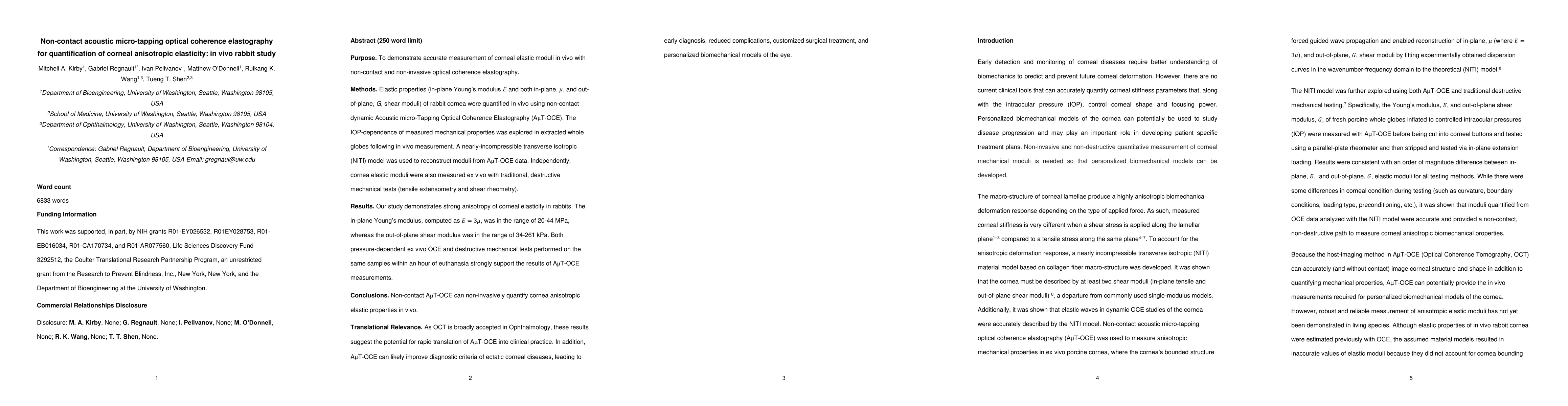
Purpose. To demonstrate accurate measurement of corneal elastic moduli in vivo with non-contact and non-invasive optical coherence elastography. Methods. Elastic properties (in-plane Young's modulus E and both in-plane, u, and out-of-plane, G, shear moduli) of rabbit cornea were quantified in vivo using non-contact dynamic Acoustic micro-Tapping Optical Coherence Elastography (AuT-OCE). The IOP-dependence of measured mechanical properties was explored in extracted whole globes following in vivo measurement. A nearly-incompressible transverse isotropic (NITI) model was used to reconstruct moduli from AuT-OCE data. Independently, cornea elastic moduli were also measured ex vivo with traditional, destructive mechanical tests (tensile extensometry and shear rheometry). Results. Our study demonstrates strong anisotropy of corneal elasticity in rabbits. The in-plane Young's modulus, computer as E=3u, was in the range of 20-44 MPa, whereas the out-of-plane shear modulus was in the range of 34-261 kPa. Both pressure-dependent ex vivo OCE and destructive mechanical tests performed on the same samples within an hour of euthanasia strongly support the results of AuT-OCE measurements. Conclusions. Non-contact AuT-OCE can non-invasively quantify cornea anisotropic elastic properties in vivo. Translational Relevance. As OCT is broadly accepted in Ophthalmology, these results suggest the potential for rapid translation of AuT-OCE into clinical practice. In addition, AuT-OCE can likely improve diagnostic criteria of ectatic corneal diseases, leading to early diagnosis, reduced complications, customized surgical treatment, and personalized biomechanical models of the eye.
AI Key Findings
Get AI-generated insights about this paper's methodology, results, significance, and more — seven facets brought into focus.
Impact
Paper Details
Authors
PDF Preview
Key Terms
Citation Network
Current paper (gray), citations (green), references (blue)
Display is limited for performance on very large graphs.
Discussion 0