Summary
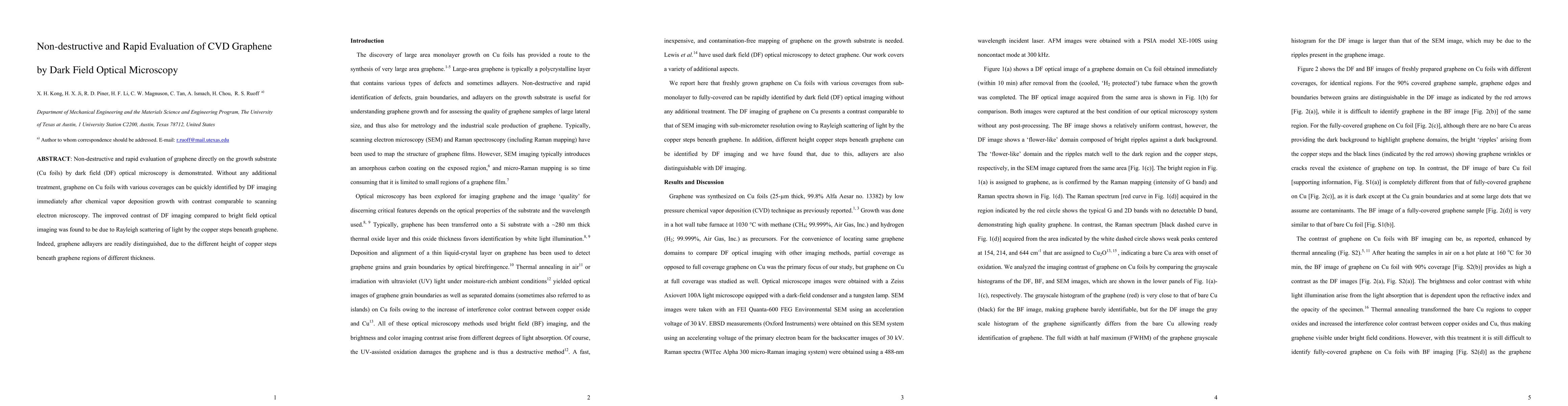
Non-destructive and rapid evaluation of graphene directly on the growth substrate (Cu foils) by dark field (DF) optical microscopy is demonstrated. Without any additional treatment, graphene on Cu foils with various coverages can be quickly identified by DF imaging immediately after chemical vapor deposition growth with contrast comparable to scanning electron microscopy. The improved contrast of DF imaging compared to bright field optical imaging was found to be due to Rayleigh scattering of light by the copper steps beneath graphene. Indeed, graphene adlayers are readily distinguished, due to the different height of copper steps beneath graphene regions of different thickness.
AI Key Findings
Get AI-generated insights about this paper's methodology, results, and significance.
Paper Details
PDF Preview
Key Terms
Citation Network
Current paper (gray), citations (green), references (blue)
Display is limited for performance on very large graphs.
| Title | Authors | Year | Actions |
|---|
Comments (0)