Publication

Published: Jun 27, 2007
Categories: cond-mat.other

Metrics

Source: ArXiv

Quick Actions

View PDF

Paper Preview

Abstract

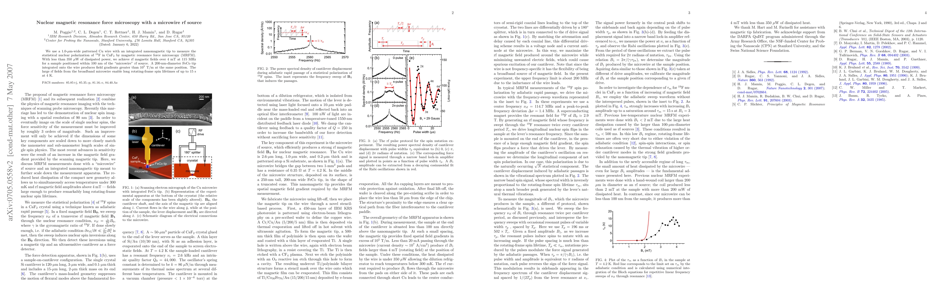
We use a 1.0-um-wide patterned Cu wire with an integrated nanomagnetic tip to measure the statistical nuclear polarization of 19F in CaF2 by magnetic resonance force microscopy (MRFM). With less than 350 uW of dissipated power, we achieve rf magnetic fields over 4 mT at 115 MHz for a sample positioned within 100 nm of the "microwire" rf source. A 200-nm diameter FeCo tip integrated onto the wire produces field gradients greater than 10^5 T/m at the same position. The large rf fields from the broadband microwire enable long rotating-frame spin lifetimes of up to 15 s at 4 K.

AI Key Findings

Get AI-generated insights about this paper's methodology, results, and significance.

Paper Details

Paper ID: 0705.0558
Journal: Appl. Phys. Lett. 90, 263111 (2007)
Comments: 4 pages, 4 figures
Categories:
cond-mat.other

How to Cite This Paper

BibTeX
@article{anon2007nuclear,
  title         = {Nuclear magnetic resonance force microscopy with a microwire rf source},
  year          = {2007},
  eprint        = {0705.0558},
  archivePrefix = {arXiv},
  primaryClass  = {cond-mat.other},
  doi           = {10.1063/1.2752536},
}
APA 7
(2007). Nuclear magnetic resonance force microscopy with a microwire rf source. arXiv. https://doi.org/10.1063/1.2752536
MLA 9
"Nuclear magnetic resonance force microscopy with a microwire rf source." arXiv, 2007, doi.org/10.1063/1.2752536.

Key Terms

rf (0.364)
wire (0.310)
tip (0.303)
nm (0.248)
integrated (0.226)
cu (0.175)
lifetimes (0.173)
fields (0.171)

Citation Network

Current paper (gray), citations (green), references (blue)

Display is limited for performance on very large graphs.

Current Paper
Citations
References
Click to view
Title Authors Year Actions

Comments (0)

This website uses cookies to ensure you get the best experience. Learn more