Mitotic activity is an important feature for grading several cancer types.
Counting mitotic figures (MFs) is a time-consuming, laborious task prone to
inter-observer variation. Inaccurate recognition of MFs can lead to incorrect
grading and hence potential suboptimal treatment. In this study, we propose an
artificial intelligence (AI)-aided approach to detect MFs in digitised
haematoxylin and eosin-stained whole slide images (WSIs). Advances in this area
are hampered by the limited number and types of cancer datasets of MFs. Here we
establish the largest pan-cancer dataset of mitotic figures by combining an
in-house dataset of soft tissue tumours (STMF) with five open-source mitotic
datasets comprising multiple human cancers and canine specimens (ICPR, TUPAC,
CCMCT, CMC and MIDOG++). This new dataset identifies 74,620 MFs and 105,538
mitotic-like figures. We then employed a two-stage framework (the Optimised
Mitoses Generator Network (OMG-Net) to classify MFs. The framework first
deploys the Segment Anything Model (SAM) to automate the contouring of MFs and
surrounding objects. An adapted ResNet18 is subsequently trained to classify
MFs. OMG-Net reaches an F1-score of 0.84 on pan-cancer MF detection (breast
carcinoma, neuroendocrine tumour and melanoma), largely outperforming the
previous state-of-the-art MIDOG++ benchmark model on its hold-out testing set
(e.g. +16% F1-score on breast cancer detection, p<0.001) thereby providing
superior accuracy in detecting MFs on various types of tumours obtained with
different scanners.
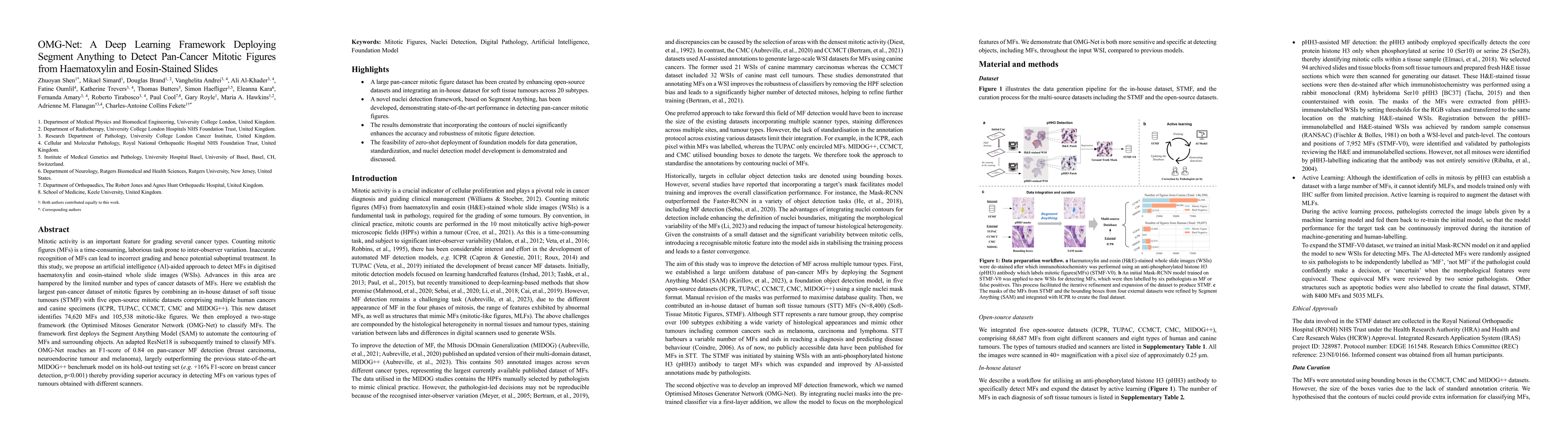
Discussion 0