Publication
Metrics
AI Quick Summary
This research introduces optical widefield NMR microscopy using NV centers in diamond to convert NMR signals into optical signals, enabling high-resolution imaging across a wide field of view. The method provides multicomponent information on signal amplitude, phase, and magnetic field properties, demonstrating potential applications in physical and life sciences.
Paper Preview
Abstract
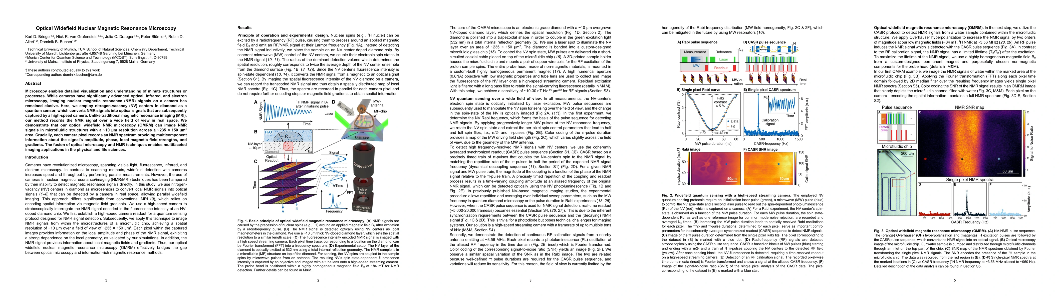
Microscopy enables detailed visualization and understanding of minute structures or processes. While cameras have significantly advanced optical, infrared, and electron microscopy, imaging nuclear magnetic resonance (NMR) signals on a camera has remained elusive. Here, we employ nitrogen-vacancy (NV) centers in diamond as a quantum sensor, which converts NMR signals into optical signals that are subsequently captured by a high-speed camera. Unlike traditional magnetic resonance imaging (MRI), our method records the NMR signal over a wide field of view in real space. We demonstrate that our optical widefield NMR microscopy (OMRM) can image NMR signals in microfluidic structures with a $\sim 10\,\mu m$ resolution across a $\sim 235 \times 150\,\mu m^2$ area. Crucially, each camera pixel records an NMR spectrum providing multicomponent information about the signal's amplitude, phase, local magnetic field strengths, and gradients. The fusion of optical microscopy and NMR techniques enables multifaceted imaging applications in the physical and life sciences.
AI Key Findings
Get AI-generated insights about this paper's methodology, results, significance, and more — seven facets brought into focus.
Impact
Paper Details
Authors
PDF Preview
Key Terms
Citation Network
Current paper (gray), citations (green), references (blue)
Display is limited for performance on very large graphs.
Discussion 0