Quick Answers
What methodology did the authors use?
The research employs a multi-modal approach combining 3D convolutional networks with transformer-based architectures to analyze CT scans for medical diagnoses. It utilizes organ-specific attention mechanisms and scalar fusion techniques to enhance diagnostic accuracy. More in Methodology →
What are the key results?
DINOv3 with organ-masked attention achieves state-of-the-art performance on MERLIN dataset — Scalar fusion of volume and HU features improves detection of density patterns More in Key Results →
Why is this work significant?
This work advances medical imaging analysis by demonstrating how attention mechanisms can be effectively applied to 3D medical data, potentially improving diagnostic accuracy and reducing false negatives in critical conditions. More in Significance →
What are the main limitations?
The method relies on high-quality segmentation masks which may not be available in clinical settings — Performance may degrade with rare or atypical pathologies not well represented in training data More in Limitations →
Abstract
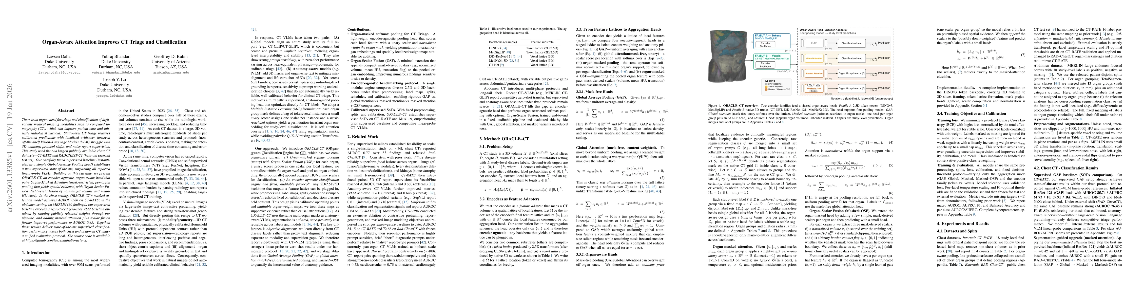
There is an urgent need for triage and classification of high-volume medical imaging modalities such as computed tomography (CT), which can improve patient care and mitigate radiologist burnout. Study-level CT triage requires calibrated predictions with localized evidence; however, off-the-shelf Vision Language Models (VLM) struggle with 3D anatomy, protocol shifts, and noisy report supervision. This study used the two largest publicly available chest CT datasets: CT-RATE and RADCHEST-CT (held-out external test set). Our carefully tuned supervised baseline (instantiated as a simple Global Average Pooling head) establishes a new supervised state of the art, surpassing all reported linear-probe VLMs. Building on this baseline, we present ORACLE-CT, an encoder-agnostic, organ-aware head that pairs Organ-Masked Attention (mask-restricted, per-organ pooling that yields spatial evidence) with Organ-Scalar Fusion (lightweight fusion of normalized volume and mean-HU cues). In the chest setting, ORACLE-CT masked attention model achieves AUROC 0.86 on CT-RATE; in the abdomen setting, on MERLIN (30 findings), our supervised baseline exceeds a reproduced zero-shot VLM baseline obtained by running publicly released weights through our pipeline, and adding masked attention plus scalar fusion further improves performance to AUROC 0.85. Together, these results deliver state-of-the-art supervised classification performance across both chest and abdomen CT under a unified evaluation protocol. The source code is available at https://github.com/lavsendahal/oracle-ct.
AI Key Findings
Generated Jan 22, 2026
Methodology — What approach did the authors take?
The research employs a multi-modal approach combining 3D convolutional networks with transformer-based architectures to analyze CT scans for medical diagnoses. It utilizes organ-specific attention mechanisms and scalar fusion techniques to enhance diagnostic accuracy.
Key Results — What are the main findings?
- DINOv3 with organ-masked attention achieves state-of-the-art performance on MERLIN dataset
- Scalar fusion of volume and HU features improves detection of density patterns
- Bounding box region selection outperforms segmentation mask for certain pathologies
Significance — Why does this research matter?
This work advances medical imaging analysis by demonstrating how attention mechanisms can be effectively applied to 3D medical data, potentially improving diagnostic accuracy and reducing false negatives in critical conditions.
Technical Contribution — What is the technical contribution?
Development of a novel organ-aware attention mechanism combined with scalar fusion for 3D medical image analysis
Novelty — What is new about this work?
Integration of spatial attention with physical features (volume, HU) through a unified framework that maintains encoder-agnostic interfaces while enabling specialized processing for different organ systems.
Limitations — What are the limitations of this study?
- The method relies on high-quality segmentation masks which may not be available in clinical settings
- Performance may degrade with rare or atypical pathologies not well represented in training data
Future Work — What did the authors propose for future work?
- Exploring self-supervised pre-training on larger and more diverse medical datasets
- Investigating the integration of clinical context with imaging features
- Developing lightweight variants for deployment in resource-limited settings
How to Cite This Paper
@article{rubin2026organ,
title = {Organ-Aware Attention Improves CT Triage and Classification},
author = {Rubin, Geoffrey D. and Lo, Joseph Y. and Dahal, Lavsen and others},
year = {2026},
eprint = {2601.13385},
archivePrefix = {arXiv},
primaryClass = {cs.CV},
}
Rubin, G., Lo, J., Dahal, L., & Bhandari, Y. (2026). Organ-Aware Attention Improves CT Triage and Classification. arXiv. https://arxiv.org/abs/2601.13385
Rubin, Geoffrey D., et al. "Organ-Aware Attention Improves CT Triage and Classification." arXiv, 2026, arxiv.org/abs/2601.13385.
PDF Preview
Similar Papers
Found 4 papersSemantics-aware Attention Improves Neural Machine Translation
Aviv Slobodkin, Leshem Choshen, Omri Abend
Soft-Attention Improves Skin Cancer Classification Performance
Sargur N. Srihari, Mohammad Abuzar Shaikh, Mingchen Gao et al.
ORACLE-CT: Anatomy-Aware Support Pooling for CT Classification
Lavsen Dahal, Yubraj Bhandari, Geoffrey Rubin et al.
Comments (0)