Quick Summary:

This paper demonstrates a method to overcome contrast reversals in ptychography for thick materials, showing that slight defocus can enhance image clarity. This approach allows for efficient imaging of strongly scattering samples in STEM, revealing atomic structures more effectively.

Summary

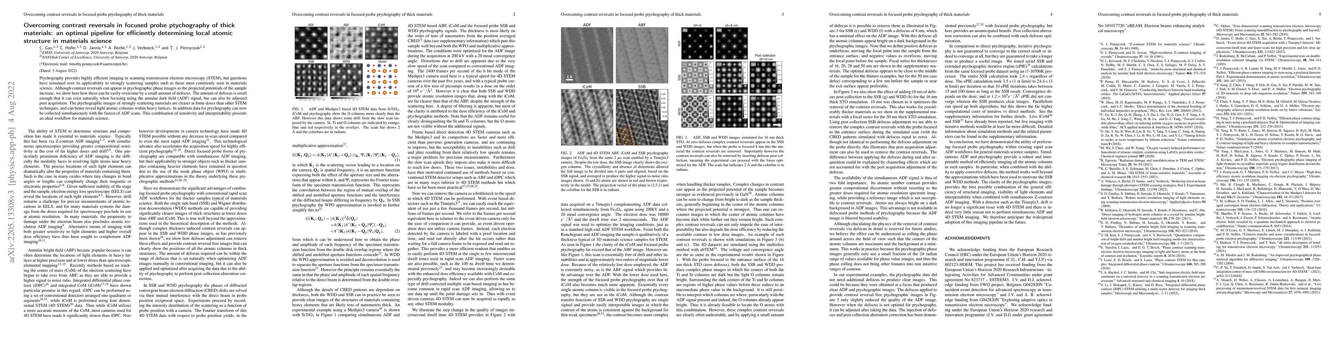
Ptychography provides highly efficient imaging in scanning transmission electron microscopy (STEM), but questions have remained over its applicability to strongly scattering samples such as those most commonly seen in materialsscience. Although contrast reversals can appear in ptychographic phase images as the projected potentials of the sample increase, we show here how these can be easily overcome by a small amount of defocus. The amount of defocus is small enough that it can exist naturally when focusing using the annular dark field (ADF) signal, but can also be adjusted post acquisition. The ptychographic images of strongly scattering materials are clearer at finite doses than other STEM techniques, and can better reveal light atomic columns within heavy lattices. In addition data for ptychography can now be collected simultaneously with the fastest of ADF scans. This combination of sensitivity and interpretability presents an ideal workflow for materials science.

AI Key Findings

Get AI-generated insights about this paper's methodology, results, and significance.

Paper Details

PDF Preview

Download PDF

Key Terms

small (0.157)
appear (0.133)
presents (0.125)
efficient (0.105)
finite (0.093)
phase (0.091)
data (0.069)
using (0.063)

Citation Network

Current paper (gray), citations (green), references (blue)

Current Paper
Citations
References
Click to view
Title Authors Year Actions
Near-Isotropic Sub-{\AA}ngstrom 3D Resolution Phase Contrast Imaging Achieved by End-to-End Ptychographic Electron Tomography
Shengboy You, Andrey Romanov, Philipp Pelz
2024
View Paper
Local-orbital ptychography for ultrahigh-resolution imaging.
Wen-Yun Yang, Haozhi Sha, Jizhe Cui, Liangze Mao, Rong Yu
2024
View Paper
Phase offset method of ptychographic contrast reversal correction.
C. Hofer, Chuang Gao, Tamazouzt Chennit, Biao Yuan, T. Pennycook
2023
View Paper
Reliable phase quantification in focused probe electron ptychography of thin materials.
C. Hofer, T. Pennycook
2023
View Paper
Resolution of Virtual Depth Sectioning from Four-Dimensional Scanning Transmission Electron Microscopy.
E. Terzoudis-Lumsden, T. Petersen, H. Brown, P. Pelz, C. Ophus, S. Findlay
2023
View Paper
On central focusing for contrast optimization in direct electron ptychography of thick samples.
C. Gao, C. Hofer, T. Pennycook
2023
View Paper
Atomic Resolution Cryogenic 4D-STEM Imaging via Robust Distortion Correction.
Jacob G. Smith, Zhennan Huang, Wenpei Gao, Guannan Zhang, M. Chi
2023
View Paper
Determining the Projected Crystal Structure from Four-dimensional Scanning Transmission Electron Microscopy via the Scattering Matrix.
A. Sadri, S. Findlay
2023
View Paper
Ptychographic measurements of varying size and shape along zeolite channels
Haozhi Sha, Jizhe Cui, Jialu Li, Yuxuan Zhang, Wen-Yun Yang, Yadong Li, Rong Yu
2023
View Paper
Detecting charge transfer at defects in 2D materials with electron ptychography.
C. Hofer, Jacob Madsen, T. Susi, T. Pennycook
2023
View Paper
Picometer-precision few-tilt ptychotomography of 2D materials
C. Hofer, K. Mustonen, V. Skákalová, T. Pennycook
2021
View Paper
Detecting charge transfer in defective 2D WS 2 with electron ptychography
C. Hofer, J. Madson, T. Susi, T. Pennycook
2023
View Paper

Comments (0)

This website uses cookies to ensure you get the best experience. Learn more