Summary
Photon Absorption Remote Sensing (PARS) enables label-free imaging of subcellular morphology by observing biomolecule specific absorption interactions. Coupled with deep-learning, PARS produces label-free virtual Hematoxylin and Eosin (H&E) stained images in unprocessed tissues. This study evaluates the diagnostic performance of these PARS-derived virtual H&E images in benign and malignant excisional skin biopsies, including Squamous (SCC), Basal (BCC) Cell Carcinoma, and normal skin. Sixteen unstained formalin-fixed paraffin-embedded skin excisions were PARS imaged, virtually H&E stained, then chemically stained and imaged at 40x. Seven fellowship trained dermatopathologists assessed all 32 images in a masked randomized fashion. Concordance analysis indicates 95.5% agreement between primary diagnoses rendered on PARS versus H&E images (Cohen's k=0.93). Inter-rater reliability was near-perfect for both image types (Fleiss' k=0.89 for PARS, k=0.80 for H&E). For subtype classification, agreement was near-perfect 91% (k=0.73) for SCC and was perfect for BCC. When assessing malignancy confinement (e.g., cancer margins), agreement was 92% between PARS and H&E (k=0.718). During assessment dermatopathologists could not reliably distinguish image origin (PARS vs. H&E), and diagnostic confidence was equivalent between the modalities. Inter-rater reliability for PARS virtual H&E was consistent with reported benchmarks for histologic evaluation. These results indicate that PARS virtual histology may be diagnostically equivalent to traditional H&E staining in dermatopathology diagnostics, while enabling assessment directly from unlabeled, or unprocessed slides. In turn, the label-free PARS virtual H&E imaging workflow may preserve tissue for downstream analysis while producing data well-suited for AI integration potentially accelerating and enhancing the accuracy of skin cancer diagnostics.
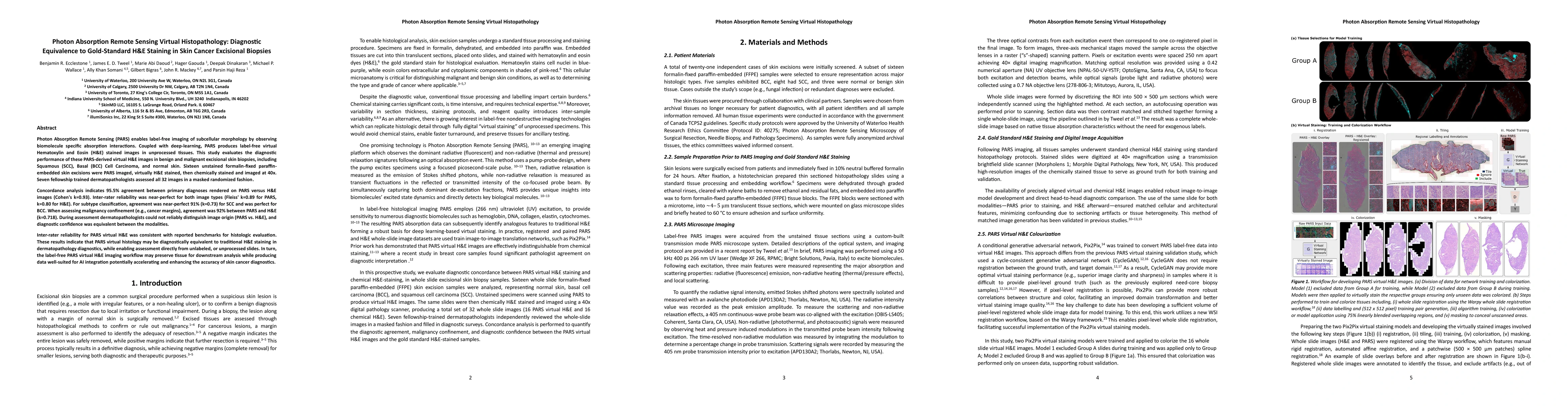
AI Key Findings
Generated Jun 09, 2025
Methodology
This study evaluated the diagnostic performance of Photon Absorption Remote Sensing (PARS) generated virtual H&E images compared to conventional chemical H&E staining in excised skin tissue specimens. Sixteen unstained FFPE skin excisional biopsies were imaged using PARS, virtually stained, then chemically labelled with H&E and digitally scanned at 40× magnification. The resulting 32 images were assessed by seven fellowship-trained dermatopathologists in a masked and randomized fashion.
Key Results
- PARS images demonstrated significant concordance (>90% agreement, with a κ >0.7) comparable to, or exceeding, chemical H&E evaluation of the same tissue section.
- Inter-rater agreement was near-perfect for both PARS (Fleiss’ kappa 𝜅=0.886) and H&E images (Fleiss’ kappa 𝜅=0.805).
- Inter-modality agreement was even stronger between the PARS and H&E images with a total 95.5% agreement (Cohen’s kappa 𝜅=0.93).
- Substantial concordance was achieved for malignancy subtype or grade, including 91% inter-modality agreement (κ=0.73) for SCC subtyping.
- Pathologists interpreted PARS images with similar confidence and accuracy and could not reliably distinguish image origin.
Significance
This research suggests that PARS virtual histology in skin excisions is diagnostically equivalent to chemical H&E staining, potentially enabling more consistent diagnosis by reducing variability associated with chemical protocols.
Technical Contribution
PARS label-free virtual histology imaging offers a promising alternative to chemical staining workflows, generating rich datasets for AI imaging and diagnostic methods.
Novelty
This work demonstrates that PARS virtual H&E imaging provides diagnostic performance equivalent to conventional H&E, with potential for enhancing diagnostic processes through standardization and AI integration.
Limitations
- The study is limited by a small sample size of tissues (n=16 slides) and pathologists (n=7 raters).
- The diagnostic scope is limited to common skin lesions (BCC and SCC), restricting generalization to other cancer subtypes or tissue types.
Future Work
- Expand the number of raters and increase the number of samples to further generalize the findings.
- Include additional histochemical stains and explore multiplexed virtual staining contrasts from a single PARS scan.
Paper Details
PDF Preview
Citation Network
Current paper (gray), citations (green), references (blue)
Display is limited for performance on very large graphs.
Similar Papers
Found 4 papersMulti-Channel Feature Extraction for Virtual Histological Staining of Photon Absorption Remote Sensing Images
Paul Fieguth, Benjamin R. Ecclestone, James E. D. Tweel et al.
Virtual Histological Staining of Label-Free Total Absorption Photoacoustic Remote Sensing (TA-PARS)
John R. Mackey, Paul Fieguth, Parsin Haji Reza et al.
Virtual Histology with Photon Absorption Remote Sensing using a Cycle-Consistent Generative Adversarial Network with Weakly Registered Pairs
Paul Fieguth, Benjamin R. Ecclestone, James E. D. Tweel et al.
No citations found for this paper.
Comments (0)