Publication
Metrics
AI Quick Summary
The paper proposes a physics-based reward function coupled with Bayesian Optimization to dynamically optimize image analysis workflows in electron microscopy, specifically applied to $(Y, Dy)Ba_2Cu_3O_{7-\delta}$ thin-films. The optimized Laplacian-of-Gaussian method, termed LoG*, performs better than DCNN segmentation in noisy conditions, demonstrating faster and more cost-effective analysis aligned with experimental objectives.
Paper Preview
Abstract
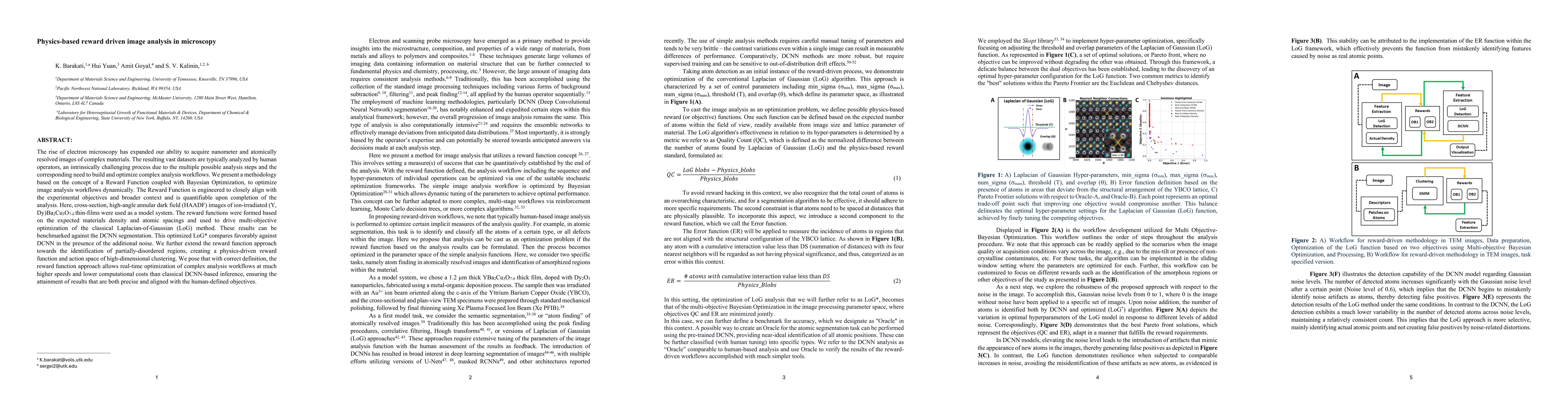
The rise of electron microscopy has expanded our ability to acquire nanometer and atomically resolved images of complex materials. The resulting vast datasets are typically analyzed by human operators, an intrinsically challenging process due to the multiple possible analysis steps and the corresponding need to build and optimize complex analysis workflows. We present a methodology based on the concept of a Reward Function coupled with Bayesian Optimization, to optimize image analysis workflows dynamically. The Reward Function is engineered to closely align with the experimental objectives and broader context and is quantifiable upon completion of the analysis. Here, cross-section, high-angle annular dark field (HAADF) images of ion-irradiated $(Y, Dy)Ba_2Cu_3O_{7-\delta}$ thin-films were used as a model system. The reward functions were formed based on the expected materials density and atomic spacings and used to drive multi-objective optimization of the classical Laplacian-of-Gaussian (LoG) method. These results can be benchmarked against the DCNN segmentation. This optimized LoG* compares favorably against DCNN in the presence of the additional noise. We further extend the reward function approach towards the identification of partially-disordered regions, creating a physics-driven reward function and action space of high-dimensional clustering. We pose that with correct definition, the reward function approach allows real-time optimization of complex analysis workflows at much higher speeds and lower computational costs than classical DCNN-based inference, ensuring the attainment of results that are both precise and aligned with the human-defined objectives.
AI Key Findings
Get AI-generated insights about this paper's methodology, results, significance, and more — seven facets brought into focus.
Impact
Paper Details
Authors
PDF Preview
Key Terms
Citation Network
Current paper (gray), citations (green), references (blue)
Display is limited for performance on very large graphs.
Discussion 0