Physics-informed motion registration of lung parenchyma across static CT images
Publication
Metrics
AI Quick Summary
This study introduces a finite element method to estimate lung strains from static CT images taken at end-expiration and end-inspiration, allowing estimation of regional deformations in lung parenchyma without requiring a temporal series of images. The method shows agreement with non-rigid image registration results using dynamic CT data.
Paper Preview
Abstract
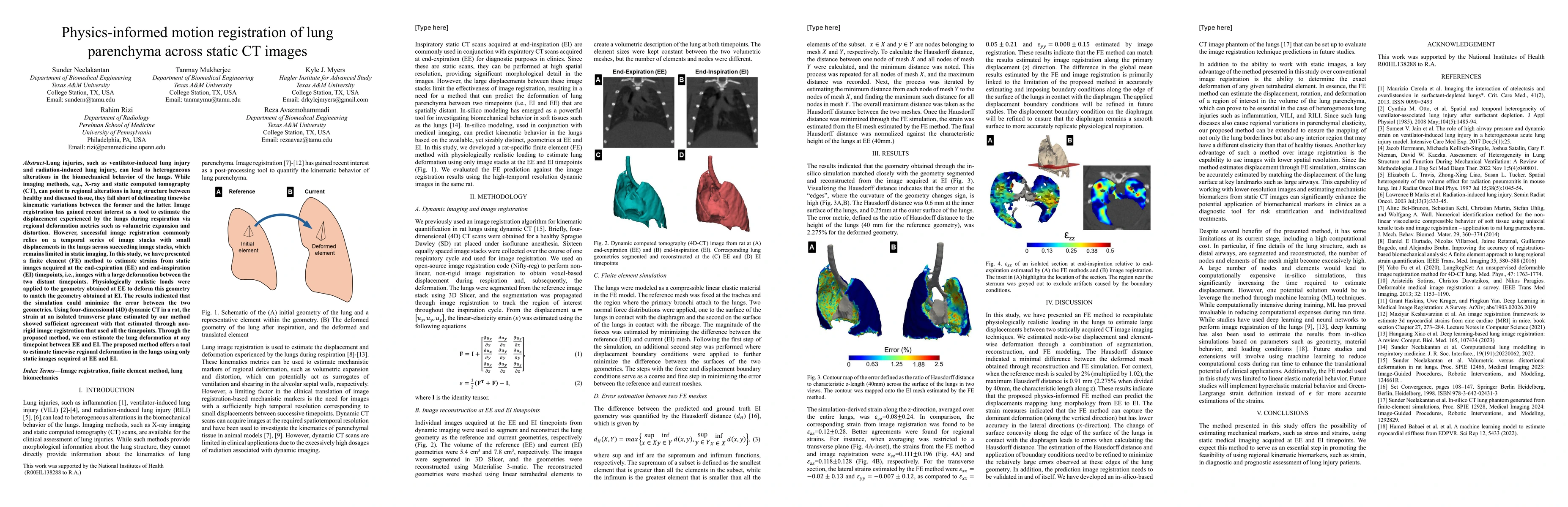
Lung injuries, such as ventilator-induced lung injury and radiation-induced lung injury, can lead to heterogeneous alterations in the biomechanical behavior of the lungs. While imaging methods, e.g., X-ray and static computed tomography (CT), can point to regional alterations in lung structure between healthy and diseased tissue, they fall short of delineating timewise kinematic variations between the former and the latter. Image registration has gained recent interest as a tool to estimate the displacement experienced by the lungs during respiration via regional deformation metrics such as volumetric expansion and distortion. However, successful image registration commonly relies on a temporal series of image stacks with small displacements in the lungs across succeeding image stacks, which remains limited in static imaging. In this study, we have presented a finite element (FE) method to estimate strains from static images acquired at the end-expiration (EE) and end-inspiration (EI) timepoints, i.e., images with a large deformation between the two distant timepoints. Physiologically realistic loads were applied to the geometry obtained at EE to deform this geometry to match the geometry obtained at EI. The results indicated that the simulation could minimize the error between the two geometries. Using four-dimensional (4D) dynamic CT in a rat, the strain at an isolated transverse plane estimated by our method showed sufficient agreement with that estimated through non-rigid image registration that used all the timepoints. Through the proposed method, we can estimate the lung deformation at any timepoint between EE and EI. The proposed method offers a tool to estimate timewise regional deformation in the lungs using only static images acquired at EE and EI.
AI Key Findings
Get AI-generated insights about this paper's methodology, results, significance, and more — seven facets brought into focus.
Impact
Paper Details
Authors
PDF Preview
Citation Network
Current paper (gray), citations (green), references (blue)
Display is limited for performance on very large graphs.
Discussion 0