This paper quantitatively analyzes various patch-based fully convolutional neural networks (FCNN) for brain tissue segmentation in MRI, comparing 2D and 3D approaches and the impact of multiple modalities and overlapping sampling strategies. The study evaluates eight FCNN architectures on diverse datasets to identify architectural strengths and weaknesses.
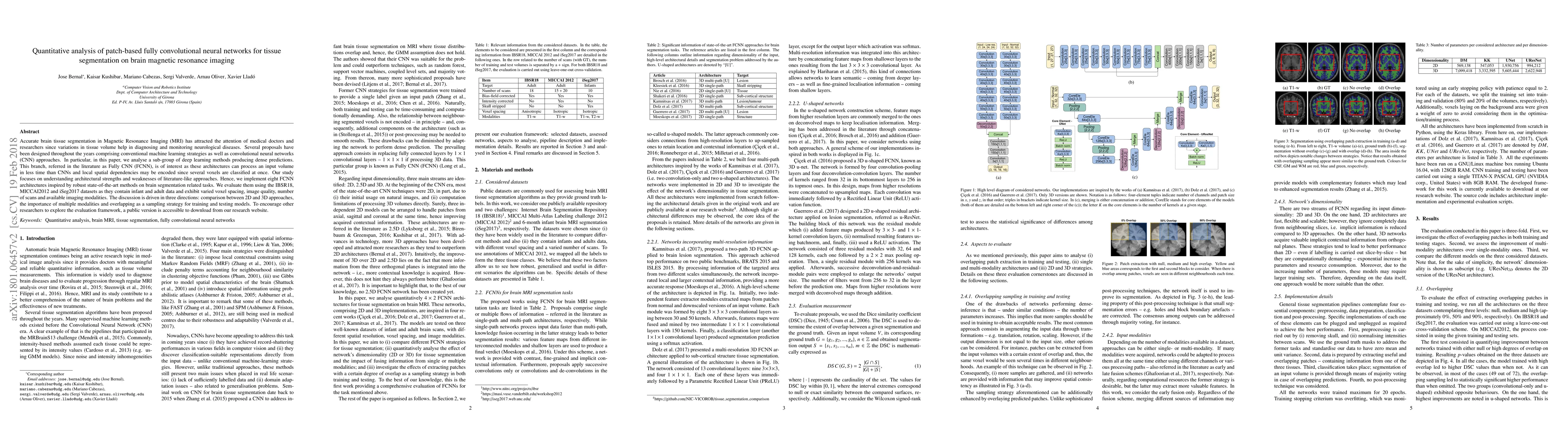
Accurate brain tissue segmentation in Magnetic Resonance Imaging (MRI) has attracted the attention of medical doctors and researchers since variations in tissue volume help in diagnosing and monitoring neurological diseases. Several proposals have been designed throughout the years comprising conventional machine learning strategies as well as convolutional neural networks (CNN) approaches. In particular, in this paper, we analyse a sub-group of deep learning methods producing dense predictions. This branch, referred in the literature as Fully CNN (FCNN), is of interest as these architectures can process an input volume in less time than CNNs and local spatial dependencies may be encoded since several voxels are classified at once. Our study focuses on understanding architectural strengths and weaknesses of literature-like approaches. Hence, we implement eight FCNN architectures inspired by robust state-of-the-art methods on brain segmentation related tasks. We evaluate them using the IBSR18, MICCAI2012 and iSeg2017 datasets as they contain infant and adult data and exhibit varied voxel spacing, image quality, number of scans and available imaging modalities. The discussion is driven in three directions: comparison between 2D and 3D approaches, the importance of multiple modalities and overlapping as a sampling strategy for training and testing models. To encourage other researchers to explore the evaluation framework, a public version is accessible to download from our research website.
This graph shows how this paper is connected to other research. The current paper is shown in dark gray, papers that cite it are in green, and papers it references are in blue. You can:
• Hover over any node to see the paper title
• Click a node to view that paper's details
• Drag nodes to rearrange the graph
• Use the zoom controls (bottom right) to zoom in/out
Title | Authors | Year | Actions |
---|---|---|---|
Enhanced U-Net for Infant Brain MRI Segmentation: A (2+1)D Convolutional Approach
|
Lehel Dénes-Fazakas, L. Kovács, György Eigner, László Szilágyi
|
2025
|
View Paper |
A comparative analysis of the health monitoring process using deep learning methods for brain tumour
|
N. Manjunathan, N. Gomathi
|
2025
|
View Paper |
Multi-atlas subcortical segmentation: an orchestration of 3D fully convolutional network and generalized mixture function
|
Jiong Wu, Shuan He, Shuang Zhou
|
2023
|
View Paper |
Role of Deep Learning in Classification of Brain MRI Images for Prediction of Disorders: A Survey of Emerging Trends
|
Poonam Rani Verma, A. Bhandari
|
2023
|
View Paper |
hist2RNA: An Efficient Deep Learning Architecture to Predict Gene Expression from Breast Cancer Histopathology Images
|
Raktim Kumar Mondol, Ewan K. A. Millar, P. Graham, L. Browne, A. Sowmya, E. Meijering
|
2023
|
View Paper |
Machine learning and deep learning in medicine and neuroimaging
|
Iván Sánchez Fernández, J. Peters
|
2023
|
View Paper |
patchIT: A Multipurpose Patch Creation Tool for Image Processing Applications
|
A. Kesidis, Vassilios Krassanakis, Loukas-Moysis Misthos, N. Merlemis
|
2022
|
View Paper |
Needle Localization and Segmentation for Radiofrequency Ablation of Liver Tumors under CT Image Guidance
|
Le Quoc Anh, Luu Manh Ha, T. van Walsum, A. Moelker, D. V. Hang, P. C. Phuong, Vu Duy Thanh
|
2022
|
View Paper |
MF2-Net: A multipath feature fusion network for medical image segmentation
|
Nagaraj Yamanakkanavar, Bumshik Lee
|
2022
|
View Paper |
Segmentation of 6-month infant brain tissues from multi-spectral MRI records using a U-Net neural network architecture
|
Lehel Dénes-Fazakas, G. Eigner, L. Szilágyi
|
2022
|
View Paper |
SVF-Net: spatial and visual feature enhancement network for brain structure segmentation
|
Qian Hu, Ying Wei, Xiang Lisa Li, Chuyuan Wang, Huan Wang, Shanze Wang
|
2022
|
View Paper |
Leveraging Deep Neural Networks to Map Caribou Lichen in High-Resolution Satellite Images Based on a Small-Scale, Noisy UAV-Derived Map
|
Shahab E. Jozdani, Dongmei Chen, Wenjun Chen, S. Leblanc, C. Prévost, Julie Lovitt, Liming He, B. Johnson
|
2021
|
View Paper |
Biomedical Image Segmentation: A Survey
|
Yahya Alzahrani, B. Boufama
|
2021
|
View Paper |
Hemorrhagic stroke lesion segmentation using a 3D U-Net with squeeze-and-excitation blocks
|
Valeriia Abramova, Albert Clérigues, A. Quiles, Deysi Garcia Figueredo, Yolanda Silva, S. Pedraza, A. Oliver, X. Lladó
|
2021
|
View Paper |
Segmentation of Brain Tissues from Infant MRI Records Using Machine Learning Techniques
|
Béla Surányi, L. Kovács, L. Szilágyi
|
2021
|
View Paper |
Generating Longitudinal Atrophy Evaluation Datasets on Brain Magnetic Resonance Images Using Convolutional Neural Networks and Segmentation Priors
|
J. Bernal, S. Valverde, Kaisar Kushibar, M. Cabezas, A. Oliver, X. Lladó
|
2021
|
View Paper |
Evaluating the Effect of Intensity Standardisation on Longitudinal Whole Brain Atrophy Quantification in Brain Magnetic Resonance Imaging
|
Emily Esmeralda Carvajal-Camelo, J. Bernal, A. Oliver, X. Lladó, M. Trujillo, The Initiative
|
2020
|
View Paper |
Convolution layer with nonlinear kernel of square of subtraction for dark-direction-free recognition of images
|
H. Yoo, Hyoseob Kim, J. Lee, Seo-Yung Lee
|
2020
|
View Paper |
Using a Patch-Wise M-Net Convolutional Neural Network for Tissue Segmentation in Brain MRI Images
|
Nagaraj Yamanakkanavar, Bumshik Lee
|
2020
|
View Paper |
MRI Segmentation and Classification of Human Brain Using Deep Learning for Diagnosis of Alzheimer’s Disease: A Survey
|
Nagaraj Yamanakkanavar, J. Choi, Bumshik Lee
|
2020
|
View Paper |
Deep Cerebellar Nuclei Segmentation via Semi-Supervised Deep Context-Aware Learning from 7T Diffusion MRI
|
Jinyoung Kim, R. Patriat, J. Kaplan, Oren Solomon, N. Harel
|
2020
|
View Paper |
ψ-Net: Stacking Densely Convolutional LSTMs for Sub-Cortical Brain Structure Segmentation
|
Lihao Liu, Xiaowei Hu, Lei Zhu, Chi-Wing Fu, J. Qin, P. Heng
|
2020
|
View Paper |
DARTS: DenseUnet-based Automatic Rapid Tool for brain Segmentation
|
Aakash Kaku, Chaitra V. Hegde, Jeffrey R. Huang, Sohae Chung, X. Wang, Matthew Young, A. Radmanesh, Y. Lui, N. Razavian
|
2019
|
View Paper |
M3Net: A multi-model, multi-size, and multi-view deep neural network for brain magnetic resonance image segmentation
|
Jie Wei, Yong Xia, Yanning Zhang
|
2019
|
View Paper |
Benchmark on Automatic Six-Month-Old Infant Brain Segmentation Algorithms: The iSeg-2017 Challenge
|
Li Wang, Dong Nie, Guannan Li, É. Puybareau, J. Dolz, Qian Zhang, Fan Wang, Jing Xia, Zhengwang Wu, Jiawei Chen, Kim-Han Thung, T. Bui, Jitae Shin, G. Zeng, Guoyan Zheng, Vladimir S Fonov, Andrew Doyle, Yongchao Xu, P. Moeskops, J. Pluim, Christian Desrosiers, Ismail Ben Ayed, G. Sanromá, O. Benkarim, A. Casamitjana, Verónica Vilaplana, Weili Lin, Gang Li, D. Shen
|
2019
|
View Paper |
Myocardial Infarction Quantification From Late Gadolinium Enhancement MRI Using Top-hat Transforms and Neural Networks
|
E. D. L. Rosa, D. Sidibé, Thomas Decourselle, T. Leclercq, A. Cochet, A. Lalande
|
2019
|
View Paper |
Asymmetric Loss Functions and Deep Densely-Connected Networks for Highly-Imbalanced Medical Image Segmentation: Application to Multiple Sclerosis Lesion Detection
|
Seyed Raein Hashemi, Seyed Sadegh Mohseni Salehi, Deniz Erdoğmuş, S. Prabhu, S. Warfield, A. Gholipour
|
2018
|
View Paper |
Tversky as a Loss Function for Highly Unbalanced Image Segmentation using 3D Fully Convolutional Deep Networks
|
Seyed Raein Hashemi, S. Salehi, Deniz Erdoğmuş, S. Prabhu, S. Warfield, A. Gholipour
|
2018
|
View Paper |
Asymmetric Similarity Loss Function to Balance Precision and Recall in Highly Unbalanced Deep Medical Image Segmentation
|
Seyed Raein Hashemi, S. Salehi, Deniz Erdoğmuş, S. Prabhu, S. Warfield, A. Gholipour
|
2018
|
View Paper |
Automated Brain Lobe Segmentation and Feature Extraction from Multiple Sclerosis Lesions Using Deep Learning
|
Nada Haj Messaoud, Rim Ayari, A. Abdallah, M. Hedi
|
2024
|
View Paper |
A Review on Alzheimer’s Disease Through Analysis of MRI Images Using Deep Learning Techniques
|
B. S. Rao, M. Aparna
|
2023
|
View Paper |
Two U-net Architectures for Infant Brain Tissue Segmentation from Multi-Spectral MRI Data
|
Lehel Dénes-Fazakas, G. Eigner, L. Kovács, László Szilágyi
|
2023
|
View Paper |
A squeeze M-SegNet architecture for segmentation of brain tissues on MRI
|
Nagaraj Yamanakkanavar, Bumshik Lee
|
2021
|
View Paper |
Automated brain segmentation methods for clinical quality MRI and CT images
|
V. Adduru
|
2020
|
View Paper |
Brain Tumor Segmentation from MRI Images Using Deep Learning Framework
|
Das Suchismita
|
2020
|
View Paper |
using Deep Learning
|
Prof. Samra Turajilic, Prof. James Larkin, Dr. Guang Yang
|
2020
|
View Paper |
Image Based Brain Segmentation: From Multi-Atlas Fusion to Deep Learning.
|
Xiangbo Lin, Xiaoxi Li
|
2019
|
View Paper |
Published: 2018-06-13
Published: 2019-07-30
Comments (0)