Towards integrating spatial localization in convolutional neural networks for brain image segmentation
Publication
Metrics
AI Quick Summary
This research explores the integration of spatial localization within convolutional neural networks (CNNs) to enhance the segmentation of brain MRIs into cerebral structures. By incorporating spatial constraints, such as distance to landmarks or a probability atlas, the study demonstrates improved consistency in segmentation predictions compared to traditional CNN approaches.
Paper Preview
Abstract
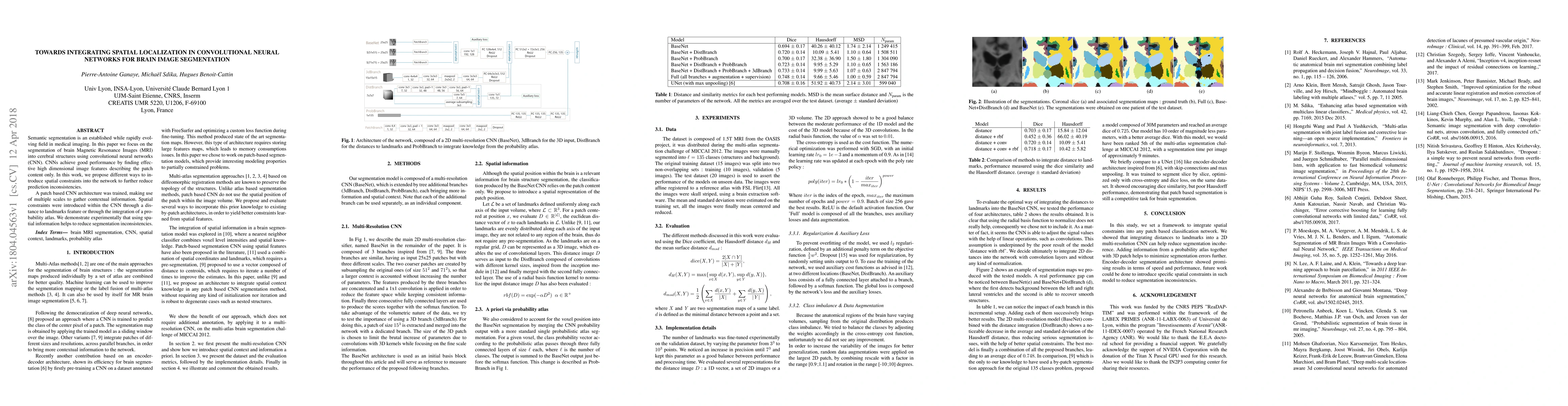
Semantic segmentation is an established while rapidly evolving field in medical imaging. In this paper we focus on the segmentation of brain Magnetic Resonance Images (MRI) into cerebral structures using convolutional neural networks (CNN). CNNs achieve good performance by finding effective high dimensional image features describing the patch content only. In this work, we propose different ways to introduce spatial constraints into the network to further reduce prediction inconsistencies. A patch based CNN architecture was trained, making use of multiple scales to gather contextual information. Spatial constraints were introduced within the CNN through a distance to landmarks feature or through the integration of a probability atlas. We demonstrate experimentally that using spatial information helps to reduce segmentation inconsistencies.
AI Key Findings
Get AI-generated insights about this paper's methodology, results, significance, and more — seven facets brought into focus.
Impact
Paper Details
PDF Preview
Key Terms
Citation Network
Current paper (gray), citations (green), references (blue)
Display is limited for performance on very large graphs.
Discussion 0