Publication
Metrics
AI Quick Summary
This paper presents the first successful X-ray diffraction imaging of single, unstained viruses, enabling visualization of their internal structure without staining. This breakthrough paves the way for quantitative imaging of a wide range of biological specimens using X-rays, including whole cells.
Paper Preview
Abstract
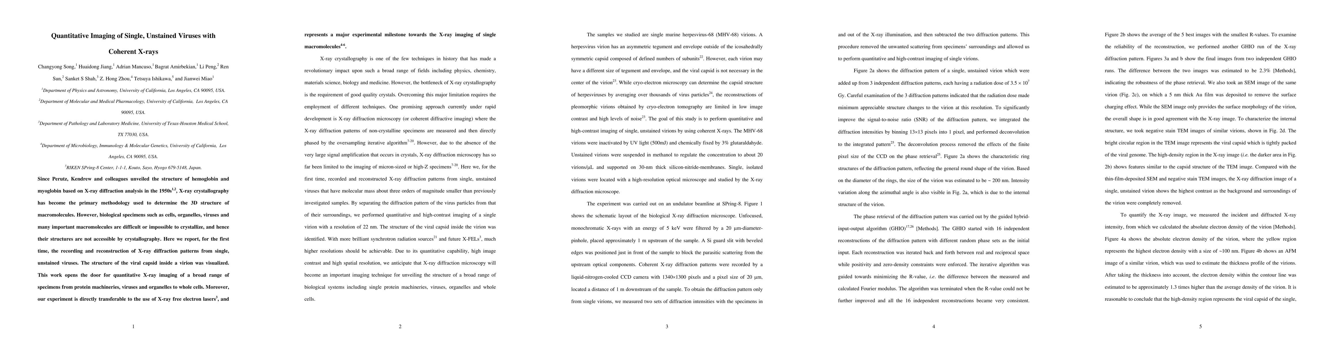
Since Perutz, Kendrew and colleagues unveiled the structure of hemoglobin and myoglobin based on X-ray diffraction analysis in the 1950s, X-ray crystallography has become the primary methodology used to determine the 3D structure of macromolecules. However, biological specimens such as cells, organelles, viruses and many important macromolecules are difficult or impossible to crystallize, and hence their structures are not accessible by crystallography. Here we report, for the first time, the recording and reconstruction of X-ray diffraction patterns from single, unstained viruses. The structure of the viral capsid inside a virion was visualized. This work opens the door for quantitative X-ray imaging of a broad range of specimens from protein machineries, viruses and organelles to whole cells. Moreover, our experiment is directly transferable to the use of X-ray free electron lasers, and represents a major experimental milestone towards the X-ray imaging of single macromolecules.
AI Key Findings
Get AI-generated insights about this paper's methodology, results, significance, and more — seven facets brought into focus.
Impact
Paper Details
PDF Preview
Key Terms
Citation Network
Current paper (gray), citations (green), references (blue)
Display is limited for performance on very large graphs.
Discussion 0