Water Window Ptychographic Imaging with Characterized Coherent X-rays
Publication
Metrics
AI Quick Summary
This paper details a ptychographic imaging experiment using focused soft X-rays at 500 eV in the water window, achieving resolutions of 53 nm for a test sample and better than 90 nm for a fossil diatom. The experiment utilized a high coherence X-ray beam and a novel non-redundant array method for coherence measurement.
Paper Preview
Abstract
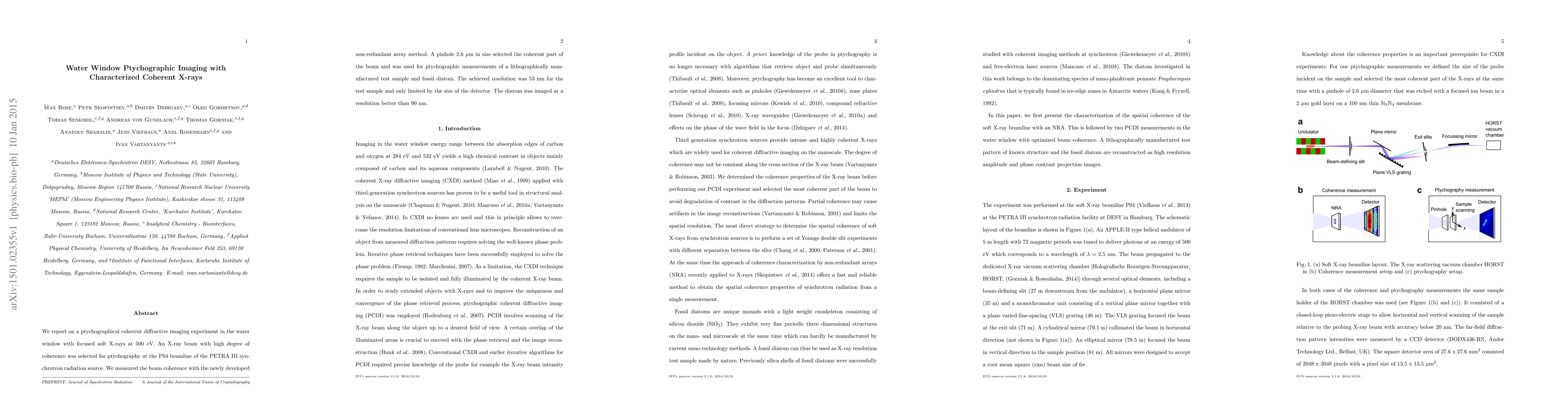
We report on a ptychographical coherent diffractive imaging experiment in the water window with focused soft X-rays at $500~\mathrm{eV}$. An X-ray beam with high degree of coherence was selected for ptychography at the P04 beamline of the PETRA III synchrotron radiation source. We measured the beam coherence with the newly developed non-redundant array method. A pinhole $2.6~\mathrm{\mu m}$ in size selected the coherent part of the beam and was used for ptychographic measurements of a lithographically manufactured test sample and fossil diatom. The achieved resolution was $53~\mathrm{nm}$ for the test sample and only limited by the size of the detector. The diatom was imaged at a resolution better than $90~\mathrm{nm}$.
AI Key Findings
Get AI-generated insights about this paper's methodology, results, significance, and more — seven facets brought into focus.
Impact
Paper Details
PDF Preview
Key Terms
Citation Network
Current paper (gray), citations (green), references (blue)
Display is limited for performance on very large graphs.
Discussion 0