Publication
Metrics
AI Quick Summary
Researchers developed a quantum magnetic resonance microscope that can image electronic spin species at high resolution, enabling imaging of tiny volumes and detecting individual spins with sensitivity of 104 spins/voxel.
Paper Preview
Abstract
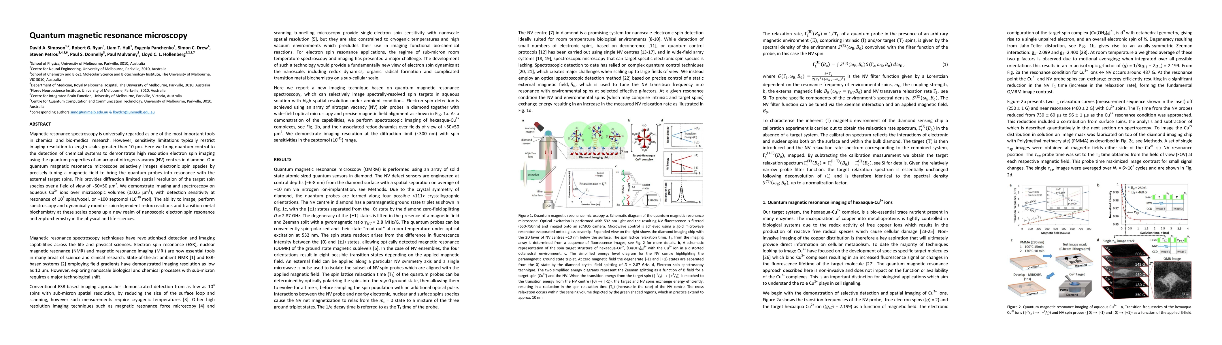
Magnetic resonance spectroscopy is universally regarded as one of the most important tools in chemical and bio-medical research. However, sensitivity limitations typically restrict imaging resolution to length scales greater than 10 \mu m. Here we bring quantum control to the detection of chemical systems to demonstrate high resolution electron spin imaging using the quantum properties of an array of nitrogen-vacancy (NV) centres in diamond. Our quantum magnetic resonance microscope selectively images electronic spin species by precisely tuning a magnetic field to bring the quantum probes into resonance with the external target spins. This provides diffraction limited spatial resolution of the target spin species over a field of view of ~50x50 \mu m^2. We demonstrate imaging and spectroscopy on aqueous Cu2+ ions over microscopic volumes (0.025 \mu m^3), with detection sensitivity at resonance of 104 spins/voxel, ~100 zeptomol (10^-19 mol). The ability to image, perform spectroscopy and dynamically monitor spin-dependent redox reactions and transition metal biochemistry at these scales opens up a new realm of nanoscopic electron spin resonance and zepto-chemistry in the physical and life sciences.
AI Key Findings
Get AI-generated insights about this paper's methodology, results, significance, and more — seven facets brought into focus.
Impact
Paper Details
PDF Preview
Key Terms
Citation Network
Current paper (gray), citations (green), references (blue)
Display is limited for performance on very large graphs.
Discussion 0