Review of speckle-tracking algorithms for x-ray phase contrast imaging: low dose applications
Publication
Metrics
AI Quick Summary
This paper reviews various algorithms for extracting phase information from near-field x-ray speckle patterns, emphasizing their applications in low-dose imaging. It discusses the advantages and limitations of each algorithm in retrieving phase-shift gradients induced by sample refraction.
Paper Preview
Abstract
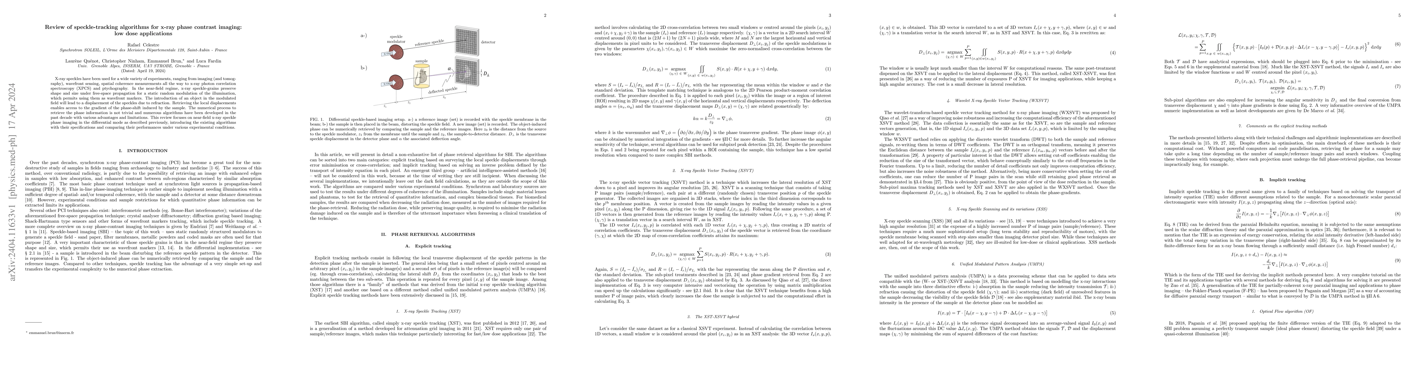
X-ray speckles have been used for a wide variety of experiments, ranging from imaging (and tomography), wavefront sensing, spatial coherence measurements all the way to x-ray photon correlation spectroscopy (XPCS) and ptychography. In the near-field regime, x-ray speckle-grains preserve shape and size under free-space propagation for a static random modulation of the illumination, which permits using them as wavefront markers. The introduction of an object in the modulated field will lead to a displacement of the speckles due to refraction. Retrieving the local displacements enables access to the gradient of the phase-shift induced by the sample. The numerical process to retrieve the phase information is not trivial and numerous algorithms have been developed in the past decade with various advantages and limitations. This review focuses on near-field x-ray speckle phase imaging in the differential mode as described previously, introducing the existing algorithms with their specifications and comparing their performances under various experimental conditions.
AI Key Findings
Get AI-generated insights about this paper's methodology, results, significance, and more — seven facets brought into focus.
Impact
Paper Details
Authors
PDF Preview
Key Terms
Citation Network
Current paper (gray), citations (green), references (blue)
Display is limited for performance on very large graphs.
Discussion 0