Authors
Publication
Metrics
Quick Actions
AI Quick Summary
This study introduces ScopeMRI, a dataset for detecting Bankart lesions on MRIs and MRAs, achieving radiologist-level diagnostic performance with deep learning models.
Quick Answers
What is "SCOPE-MRI: Bankart Lesion Detection as a Case Study in Data Curation and Deep Learning for Challenging Diagnoses" about?
This study introduces ScopeMRI, a dataset for detecting Bankart lesions on MRIs and MRAs, achieving radiologist-level diagnostic performance with deep learning models.
What methodology did the authors use?
The study introduces ScopeMRI, a publicly available, expert-annotated dataset for shoulder pathologies, and presents a deep learning framework using CNNs and transformers for detecting Bankart lesions on standard MRIs and MRAs. Models were trained, validated, and tested using sagittal, axial, and coronal view ensembles. More in Methodology →
What are the key results?
Deep learning models achieved an AUC of 0.91 and 0.93 for standard MRIs and MRAs, respectively. — Models demonstrated sensitivity of 83% and 94%, and specificity of 91% and 86% for standard MRIs and MRAs. More in Key Results →
Why is this work significant?
This research shows deep learning models can achieve radiologist-level diagnostic performance on standard MRIs, potentially reducing the need for invasive MRAs and advancing musculoskeletal imaging research. More in Significance →
What are the main limitations?
The study was limited to shoulder pathologies and did not explore other musculoskeletal conditions. — Generalizability was demonstrated across imaging protocols but further external validation is needed. More in Limitations →
Abstract
While deep learning has shown strong performance in musculoskeletal imaging, existing work has largely focused on pathologies where diagnosis is not a clinical challenge, leaving more difficult problems underexplored, such as detecting Bankart lesions (anterior-inferior glenoid labral tears) on standard MRIs. Diagnosing these lesions is challenging due to their subtle imaging features, often leading to reliance on invasive MRI arthrograms (MRAs). This study introduces ScopeMRI, the first publicly available, expert-annotated dataset for shoulder pathologies, and presents a deep learning (DL) framework for detecting Bankart lesions on both standard MRIs and MRAs. ScopeMRI includes 586 shoulder MRIs (335 standard, 251 MRAs) from 558 patients who underwent arthroscopy. Ground truth labels were derived from intraoperative findings, the gold standard for diagnosis. Separate DL models for MRAs and standard MRIs were trained using a combination of CNNs and transformers. Predictions from sagittal, axial, and coronal views were ensembled to optimize performance. The models were evaluated on a 20% hold-out test set (117 MRIs: 46 MRAs, 71 standard MRIs). The models achieved an AUC of 0.91 and 0.93, sensitivity of 83% and 94%, and specificity of 91% and 86% for standard MRIs and MRAs, respectively. Notably, model performance on non-invasive standard MRIs matched or surpassed radiologists interpreting MRAs. External validation demonstrated initial generalizability across imaging protocols. This study demonstrates that DL models can achieve radiologist-level diagnostic performance on standard MRIs, reducing the need for invasive MRAs. By releasing ScopeMRI and a modular codebase for training and evaluating deep learning models on 3D medical imaging data, we aim to accelerate research in musculoskeletal imaging and support the development of new datasets for clinically challenging diagnostic tasks.
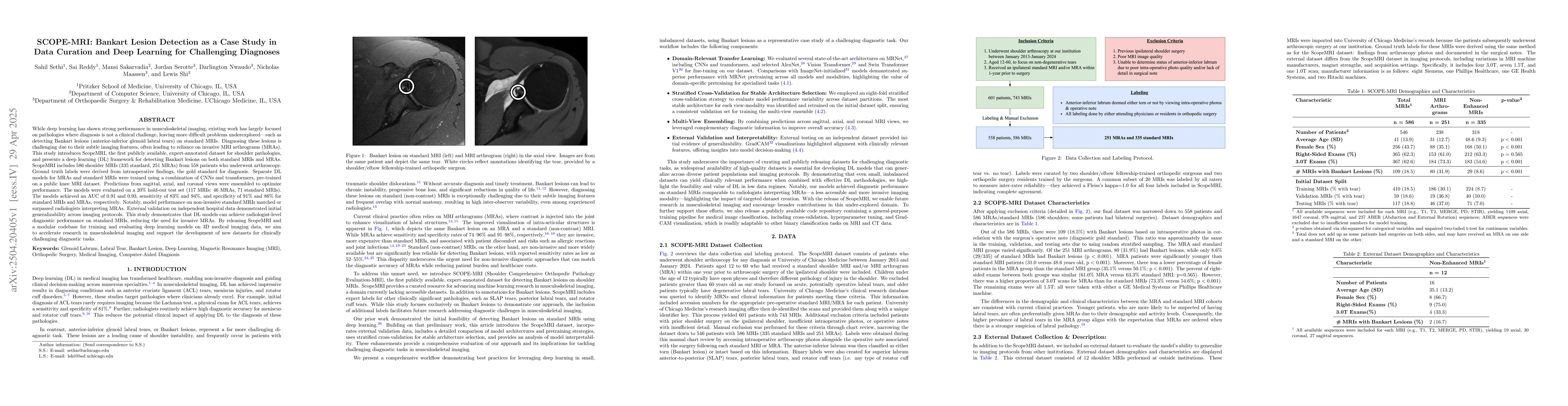
AI Key Findings
Generated Jun 08, 2025
Methodology — What approach did the authors take?
The study introduces ScopeMRI, a publicly available, expert-annotated dataset for shoulder pathologies, and presents a deep learning framework using CNNs and transformers for detecting Bankart lesions on standard MRIs and MRAs. Models were trained, validated, and tested using sagittal, axial, and coronal view ensembles.
Key Results — What are the main findings?
- Deep learning models achieved an AUC of 0.91 and 0.93 for standard MRIs and MRAs, respectively.
- Models demonstrated sensitivity of 83% and 94%, and specificity of 91% and 86% for standard MRIs and MRAs.
- Performance on non-invasive standard MRIs matched or surpassed radiologists interpreting MRAs.
Significance — Why does this research matter?
This research shows deep learning models can achieve radiologist-level diagnostic performance on standard MRIs, potentially reducing the need for invasive MRAs and advancing musculoskeletal imaging research.
Technical Contribution — What is the technical contribution?
ScopeMRI dataset and a modular codebase for training and evaluating deep learning models on 3D medical imaging data.
Novelty — What is new about this work?
First publicly available, expert-annotated dataset for shoulder pathologies, and the first to demonstrate radiologist-level diagnostic performance on non-invasive standard MRIs for Bankart lesion detection.
Limitations — What are the limitations of this study?
- The study was limited to shoulder pathologies and did not explore other musculoskeletal conditions.
- Generalizability was demonstrated across imaging protocols but further external validation is needed.
Future Work — What did the authors propose for future work?
- Explore application of the methodology to other musculoskeletal conditions.
- Conduct larger-scale external validation to confirm generalizability.
How to Cite This Paper
@article{nwaudo2025scope,
title = {SCOPE-MRI: Bankart Lesion Detection as a Case Study in Data Curation and
Deep Learning for Challenging Diagnoses},
author = {Nwaudo, Darlington and Serotte, Jordan and Shi, Lewis and others},
year = {2025},
eprint = {2504.20405},
archivePrefix = {arXiv},
primaryClass = {eess.IV},
}
Nwaudo, D., Serotte, J., Shi, L., Sakarvadia, M., Maassen, N., Sethi, S., & Reddy, S. (2025). SCOPE-MRI: Bankart Lesion Detection as a Case Study in Data Curation and
Deep Learning for Challenging Diagnoses. arXiv. https://arxiv.org/abs/2504.20405
Nwaudo, Darlington, et al. "SCOPE-MRI: Bankart Lesion Detection as a Case Study in Data Curation and
Deep Learning for Challenging Diagnoses." arXiv, 2025, arxiv.org/abs/2504.20405.
PDF Preview
Citation Network
Current paper (gray), citations (green), references (blue)
Display is limited for performance on very large graphs.
Similar Papers
Found 4 papersAutomatic Breast Lesion Detection in Ultrafast DCE-MRI Using Deep Learning
Shahriar B. Shokouhi, Jonas Teuwen, Fazael Ayatollahi et al.
Grading system for Hill-Sachs lesion and its association with Bankart lesion: a cross-sectional study.
Ahmad, Rani
Deep learning-reconstructed hepatobiliary MRI: Enhancing biliary delineation, lesion margin sharpness, and small lesion detection.
Sun, Wei, Zhang, Ming, Liu, Kai et al.
AI-Assisted MRI Interpretation in Diagnosing Bankart and Reverse Bankart Lesions.
Sethi, Sahil, Reddy, Sai, Sakarvadia, Mansi et al.
No citations found for this paper.
Comments (0)