Summary
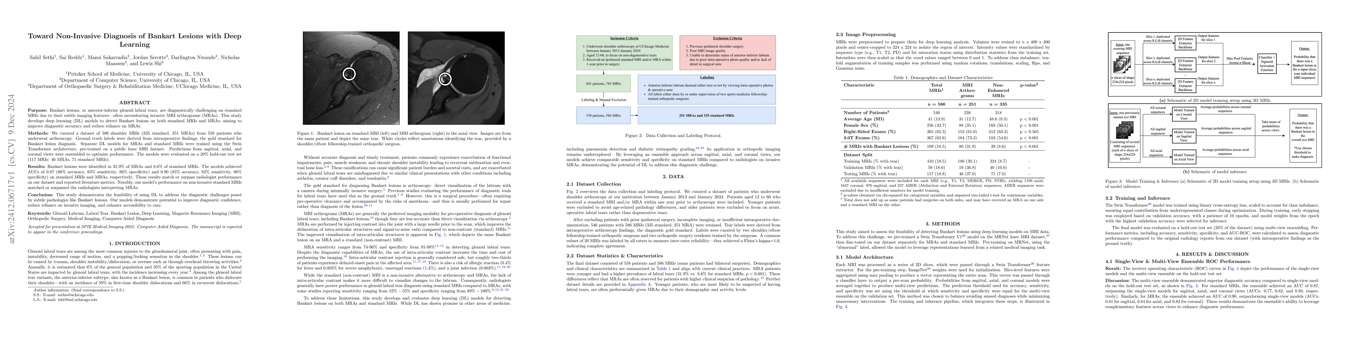
Bankart lesions, or anterior-inferior glenoid labral tears, are diagnostically challenging on standard MRIs due to their subtle imaging features-often necessitating invasive MRI arthrograms (MRAs). This study develops deep learning (DL) models to detect Bankart lesions on both standard MRIs and MRAs, aiming to improve diagnostic accuracy and reduce reliance on MRAs. We curated a dataset of 586 shoulder MRIs (335 standard, 251 MRAs) from 558 patients who underwent arthroscopy. Ground truth labels were derived from intraoperative findings, the gold standard for Bankart lesion diagnosis. Separate DL models for MRAs and standard MRIs were trained using the Swin Transformer architecture, pre-trained on a public knee MRI dataset. Predictions from sagittal, axial, and coronal views were ensembled to optimize performance. The models were evaluated on a 20% hold-out test set (117 MRIs: 46 MRAs, 71 standard MRIs). Bankart lesions were identified in 31.9% of MRAs and 8.6% of standard MRIs. The models achieved AUCs of 0.87 (86% accuracy, 83% sensitivity, 86% specificity) and 0.90 (85% accuracy, 82% sensitivity, 86% specificity) on standard MRIs and MRAs, respectively. These results match or surpass radiologist performance on our dataset and reported literature metrics. Notably, our model's performance on non-invasive standard MRIs matched or surpassed the radiologists interpreting MRAs. This study demonstrates the feasibility of using DL to address the diagnostic challenges posed by subtle pathologies like Bankart lesions. Our models demonstrate potential to improve diagnostic confidence, reduce reliance on invasive imaging, and enhance accessibility to care.
AI Key Findings
Get AI-generated insights about this paper's methodology, results, and significance.
Paper Details
PDF Preview
Citation Network
Current paper (gray), citations (green), references (blue)
Display is limited for performance on very large graphs.
Similar Papers
Found 4 papersSCOPE-MRI: Bankart Lesion Detection as a Case Study in Data Curation and Deep Learning for Challenging Diagnoses
Mansi Sakarvadia, Sahil Sethi, Sai Reddy et al.
A Review of Deep Learning Approaches for Non-Invasive Cognitive Impairment Detection
Jian Sun, Muath Alsuhaibani, Mohammad H. Mahoor et al.
| Title | Authors | Year | Actions |
|---|
Comments (0)