Seeing Cells Clearly: Evaluating Machine Vision Strategies for Microglia Centroid Detection in 3D Images
Publication
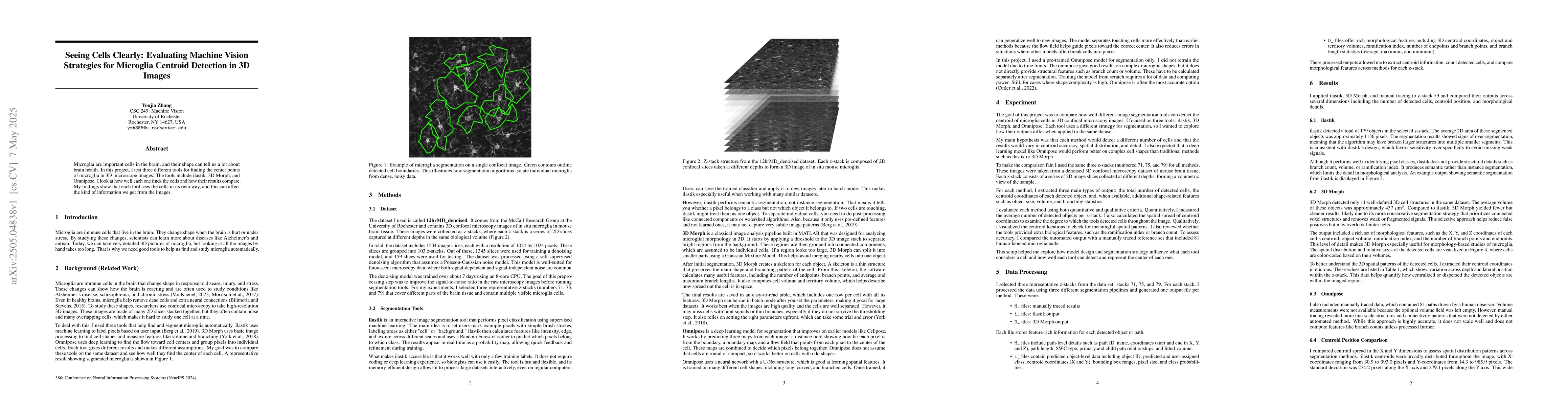
Metrics
AI Quick Summary
A study evaluated three machine vision tools for detecting microglia cells in 3D images, finding that each tool has unique strengths and limitations.
Paper Preview
Abstract
Microglia are important cells in the brain, and their shape can tell us a lot about brain health. In this project, I test three different tools for finding the center points of microglia in 3D microscope images. The tools include ilastik, 3D Morph, and Omnipose. I look at how well each one finds the cells and how their results compare. My findings show that each tool sees the cells in its own way, and this can affect the kind of information we get from the images.
AI Key Findings
Get AI-generated insights about this paper's methodology, results, significance, and more — seven facets brought into focus.
Impact
Authors
PDF Preview
Citation Network
Current paper (gray), citations (green), references (blue)
Display is limited for performance on very large graphs.
Discussion 0