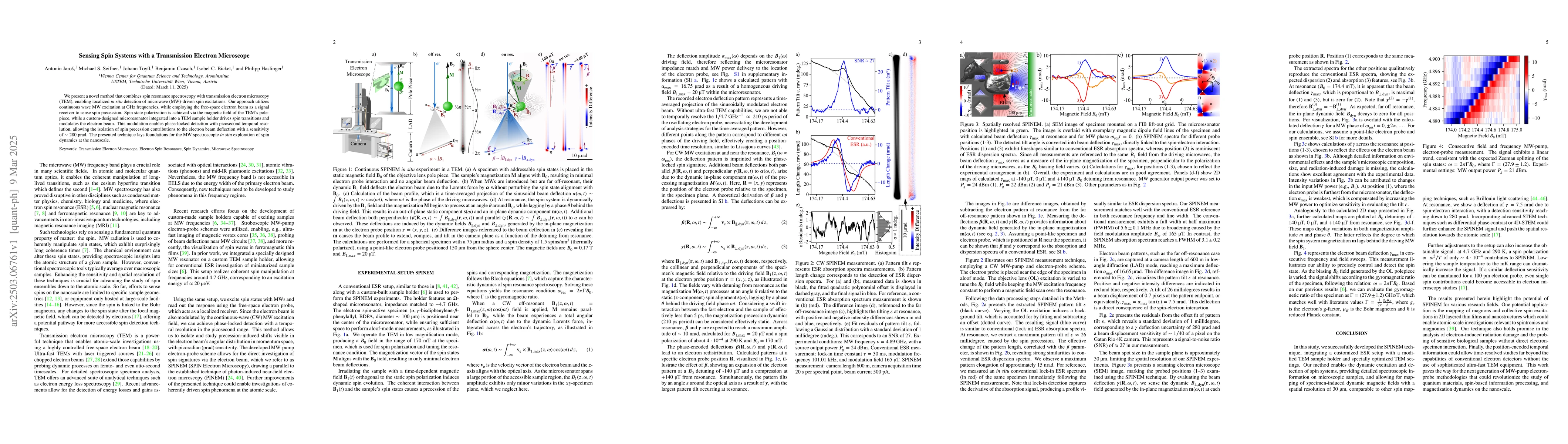
ArxivLens
ArXiv quant-ph cond-mat.mtrl-sci physics.app-ph

Sensing Spin Systems with a Transmission Electron Microscope

Antonín Jaroš, Benjamin Czasch, Isobel C. Bicket, Johann Toyfl, Michael S. Seifner, Philipp Haslinger

2503.06761 Published Mar 9, 2025
View PDF

Publication

Published: Mar 09, 2025
Categories: quant-ph, cond-mat.mtrl-sci, physics.app-ph

Metrics

Source: ArXiv

AI Quick Summary

This paper introduces a method combining spin resonance spectroscopy with TEM for in-situ detection of microwave-driven spin excitations, using GHz frequency MW excitation and a custom microresonator for spin transitions, achieving picosecond temporal resolution and a deflection sensitivity of $\sim 280$ prad. The technique enables the exploration of spin dynamics at the nanoscale.

Quick Answers

What is "Sensing Spin Systems with a Transmission Electron Microscope" about?

This paper introduces a method combining spin resonance spectroscopy with TEM for in-situ detection of microwave-driven spin excitations, using GHz frequency MW excitation and a custom microresonator for spin transitions, achieving picosecond temporal resolution and a deflection sensitivity of $\sim 280$ prad. The technique enables the exploration of spin dynamics at the nanoscale.

What methodology did the authors use?

The research presents a novel method that integrates continuous wave microwave (MW) excitation with transmission electron microscopy (TEM) to detect localized, in-situ spin excitations. This is achieved by using the TEM's polepiece magnetic field for spin state polarization and a custom-designed microresonator for driving spin transitions and modulating the electron beam, enabling phase-locked detection with picosecond temporal resolution. More in Methodology →

What are the key results?

The technique allows for the detection of spin precession contributions to electron beam deflection with a sensitivity of ~280 prad. — Experimental results align well with theoretical calculations, demonstrating the feasibility of the method for nanoscale exploration of spin dynamics. More in Key Results →

Why is this work significant?

This research is significant as it opens new avenues for nanoscale exploration of spin dynamics, potentially benefiting fields such as spintronics, magnonics, and the study of radiation damage in sensitive biological samples. More in Significance →

What are the main limitations?

The current setup has a spatial resolution of approximately 30 µm, which may limit detailed studies requiring atomic-scale precision. — Environmental effects and detailed information about the sample's microscopic composition, size, and radiation-induced damage are not provided, which could influence results in practical applications. More in Limitations →

Paper Preview

Abstract

We present a novel method that combines spin resonance spectroscopy with transmission electron microscopy (TEM), enabling localized in-situ detection of microwave (MW)-driven spin excitations. Our approach utilizes continuous wave MW excitation at GHz frequencies, while employing the free-space electron beam as a signal receiver to sense spin precession. Spin state polarization is achieved via the magnetic field of the TEM's polepiece, while a custom-designed microresonator integrated into a TEM sample holder drives spin transitions and modulates the electron beam. This modulation enables phase-locked detection with picosecond temporal resolution, allowing the isolation of spin precession contributions to the electron beam deflection with a sensitivity of $\sim 280$ prad. The presented technique lays foundations for the MW spectroscopic in-situ exploration of spin dynamics at the nanoscale.

Key Findings, in focus

Seven facets of this paper, analysed and brought into focus by AI.

AI-generated · verify against source Generated Jun 10, 2025
In focus · one-line synthesis

This research is significant as it opens new avenues for nanoscale exploration of spin dynamics, potentially benefiting fields such as spintronics, magnonics, and the study of radiation damage in sensitive biological samples.

01
MethodologyHow they did it

The research presents a novel method that integrates continuous wave microwave (MW) excitation with transmission electron microscopy (TEM) to detect localized, in-situ spin excitations. This is achieved by using the TEM's polepiece magnetic field for spin state polarization and a custom-designed microresonator for driving spin transitions and modulating the electron beam, enabling phase-locked detection with picosecond temporal resolution.

02
ResultsWhat they found
  • The technique allows for the detection of spin precession contributions to electron beam deflection with a sensitivity of ~280 prad.
  • Experimental results align well with theoretical calculations, demonstrating the feasibility of the method for nanoscale exploration of spin dynamics.
  • The method successfully distinguishes between conventional ESR absorption and dispersion spectra based on electron beam deflection patterns.
  • The approach offers spatial resolution comparable to other spin mapping techniques, with potential for further enhancement using advanced STEM techniques.
03
SignificanceWhy it matters

This research is significant as it opens new avenues for nanoscale exploration of spin dynamics, potentially benefiting fields such as spintronics, magnonics, and the study of radiation damage in sensitive biological samples.

04
Technical ContributionsWhat's new technically

The primary technical contribution is the development of the SPINEM (Spin Polarized Electron Microscopy) technique, which combines ESR with TEM for dynamic excitation and detection of spin systems, providing detailed spectroscopic information on microscopic samples and mapping specimen-induced dynamic magnetic fields with high spatial resolution.

05
NoveltyWhat sets it apart

The novelty of this work lies in its innovative integration of spin resonance spectroscopy with TEM, enabling localized, in-situ detection of MW-driven spin excitations using the electron beam as a signal receiver. This method offers a unique approach to study spin dynamics at the nanoscale with high sensitivity and temporal resolution.

06
LimitationsWhat to be cautious about
  • The current setup has a spatial resolution of approximately 30 µm, which may limit detailed studies requiring atomic-scale precision.
  • Environmental effects and detailed information about the sample's microscopic composition, size, and radiation-induced damage are not provided, which could influence results in practical applications.
  • The study does not address potential challenges in maintaining deflection sensitivity for smaller electron probes.
07
Future WorkWhere it goes next
  • Integration with advanced STEM techniques like differential phase contrast or 4D-STEM could push spatial resolution toward the atomic scale.
  • Lowering the sample temperature to the Kelvin range could dramatically increase the signal, potentially enabling the detection of single-spin contributions.
  • Further optimization of the MW-pump electron-probe setup could enhance signal strength and sensitivity.

Impact

47
References

Paper Details

Paper ID: 2503.06761
Comments: 11 pages, 5 figures
Categories:
quant-ph cond-mat.mtrl-sci physics.app-ph

How to Cite This Paper

Reference manager: RIS EndNote

Citation Network

Current paper (gray), citations (green), references (blue)

Display is limited for performance on very large graphs.

Current Paper
Citations
References
Click to view

Related Papers

4 found


Discussion 0

This website uses cookies to ensure you get the best experience. Learn more