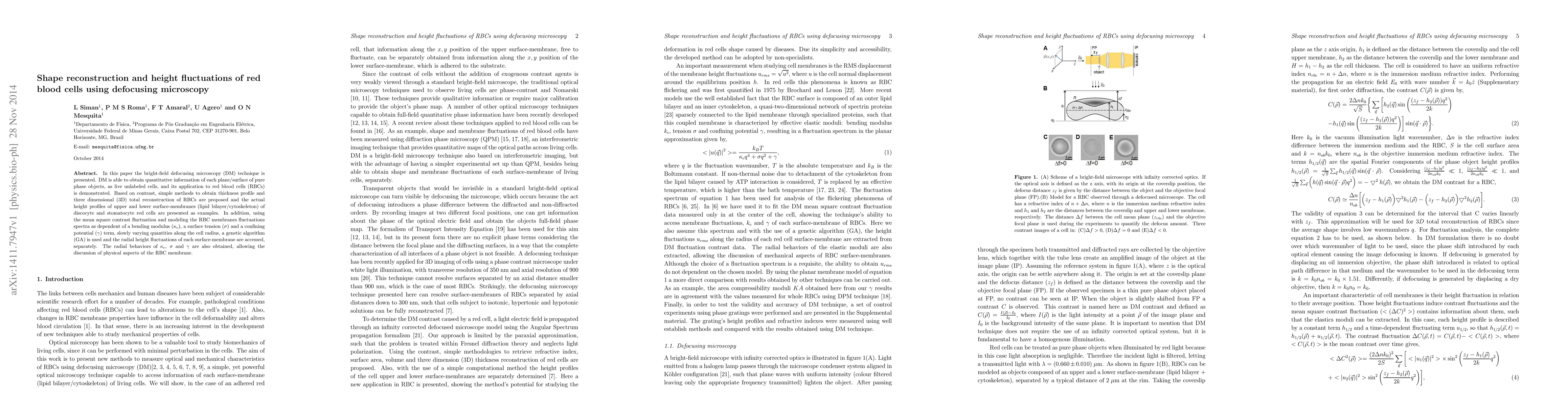
Shape reconstruction and height fluctuations of red blood cells using defocusing microscopy
Publication
Metrics
AI Quick Summary
Researchers used a microscopy technique called defocusing microscopy to study the shape and structure of red blood cells, revealing new insights into their membrane properties and height fluctuations.
Paper Preview
Abstract
In this paper the bright-field defocusing microscopy (DM) technique is presented. DM is able to obtain quantitative information of each plane/surface of pure phase objects, as live unlabeled cells, and its application to red blood cells (RBCs) is demonstrated. Based on contrast, simple methods to obtain thickness profile and three dimensional (3D) total reconstruction of RBCs are proposed and the actual height profiles of upper and lower surface-membranes (lipid bilayer$/$cytoskeleton) of discocyte and stomatocyte red cells are presented as examples. In addition, using the mean square contrast fluctuation and modeling the RBC membranes fluctuations spectra as dependent of a bending modulus $(\kappa_c)$, a surface tension $(\sigma)$ and a confining potential $(\gamma)$ term, slowly varying quantities along the cell radius, a genetic algorithm (GA) is used and the radial height fluctuations of each surface-membrane are accessed, separately. The radial behaviors of $\kappa_c$, $\sigma$ and $\gamma$ are also obtained, allowing the discussion of physical aspects of the RBC membrane.
AI Key Findings
Get AI-generated insights about this paper's methodology, results, significance, and more — seven facets brought into focus.
Impact
Paper Details
PDF Preview
Key Terms
Citation Network
Current paper (gray), citations (green), references (blue)
Display is limited for performance on very large graphs.
Discussion 0