Publication
Metrics
Paper Preview
Abstract
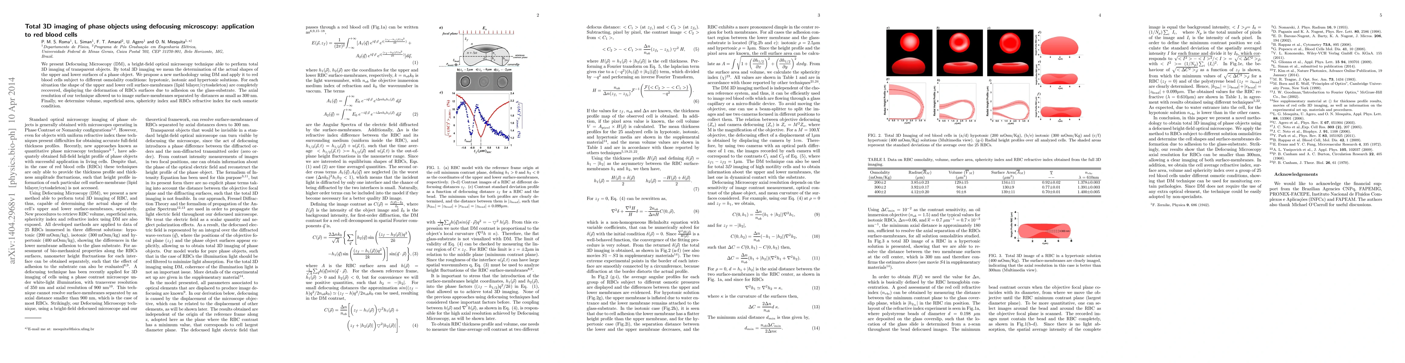
We present Defocusing Microscopy (DM), a bright-field optical microscopy technique able to perform total 3D imaging of transparent objects. By total 3D imaging we mean the determination of the actual shapes of the upper and lower surfaces of a phase object. We propose a new methodology using DM and apply it to red blood cells subject to different osmolality conditions: hypotonic, isotonic and hypertonic solutions. For each situation the shape of the upper and lower cell surface-membranes (lipid bilayer/cytoskeleton) are completely recovered, displaying the deformation of RBCs surfaces due to adhesion on the glass-substrate. The axial resolution of our technique allowed us to image surface-membranes separated by distances as small as 300 nm. Finally, we determine volume, superficial area, sphericity index and RBCs refractive index for each osmotic condition.
AI Key Findings
Get AI-generated insights about this paper's methodology, results, significance, and more — seven facets brought into focus.
Impact
Paper Details
PDF Preview
Key Terms
Citation Network
Current paper (gray), citations (green), references (blue)
Display is limited for performance on very large graphs.
Discussion 0