01
MethodologyHow they did it
Finite element simulation of breast compression using segmented MRI images
This paper develops a 3D biomechanical model to simulate breast compression during mammography, aiming to characterize patient discomfort, dose, and image quality. The model predicts tissue motion and stress, which correlate with patient discomfort, and assesses these factors under different compression techniques.
This paper develops a 3D biomechanical model to simulate breast compression during mammography, aiming to characterize patient discomfort, dose, and image quality. The model predicts tissue motion and stress, which correlate with patient discomfort, and assesses these factors under different compression techniques.
Finite element simulation of breast compression using segmented MRI images More in Methodology →
Compression force magnitude comparable to real subject data — SDNR and AGD computed for microcalcification conspicuity in tomosynthesis More in Key Results →
Improved patient comfort without affecting image quality and delivered dose More in Significance →
Assumes idealized breast shape and material properties — Limited to small breast volumes (cup sizes A and F) More in Limitations →
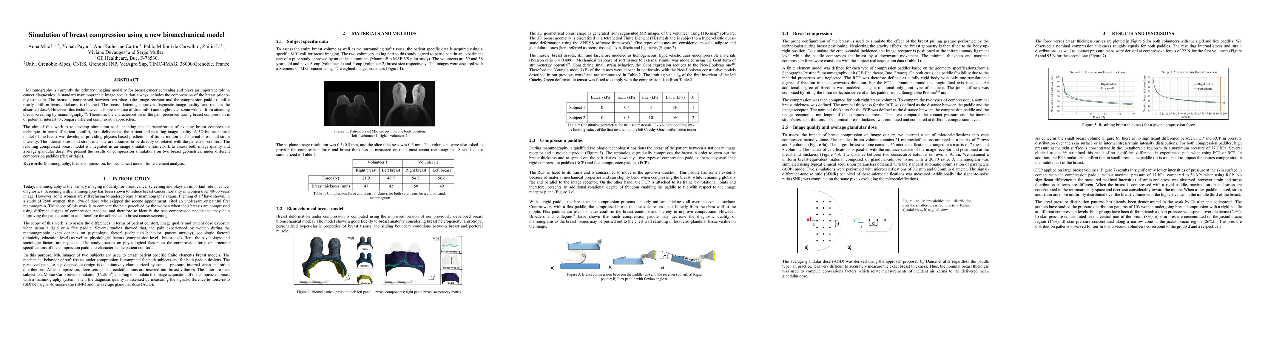
Mammography is currently the primary imaging modality for breast cancer screening and plays an important role in cancer diagnostics. A standard mammographic image acquisition always includes the compression of the breast prior x-ray exposure. The breast is compressed between two plates (the image receptor and the compression paddle) until a nearly uniform breast thickness is obtained. The breast flattening improves diagnostic image quality 1 and reduces the absorbed dose 2. However, this technique can also be a source of discomfort and might deter some women from attending breast screening by mammography 3,4. Therefore, the characterization of the pain perceived during breast compression is of potential interest to compare different compression approaches. The aim of this work is to develop simulation tools enabling the characterization of existing breast compression techniques in terms of patient comfort, dose delivered to the patient and resulting image quality. A 3D biomechanical model of the breast was developed providing physics-based predictions of tissue motion and internal stress and strain intensity. The internal stress and strain intensity are assumed to be directly correlated with the patient discomfort. The resulting compressed breast model is integrated in an image simulation framework to assess both image quality and average glandular dose. We present the results of compression simulations on two breast geometries, under different compression paddles (flex or rigid).
Seven facets of this paper, analysed and brought into focus by AI.
Improved patient comfort without affecting image quality and delivered dose
Finite element simulation of breast compression using segmented MRI images
Improved patient comfort without affecting image quality and delivered dose
Development of finite element simulation framework for breast compression optimization
Flex paddle design reduces pressure intensity without compromising image quality
Current paper (gray), citations (green), references (blue)
Display is limited for performance on very large graphs.
Discussion 0