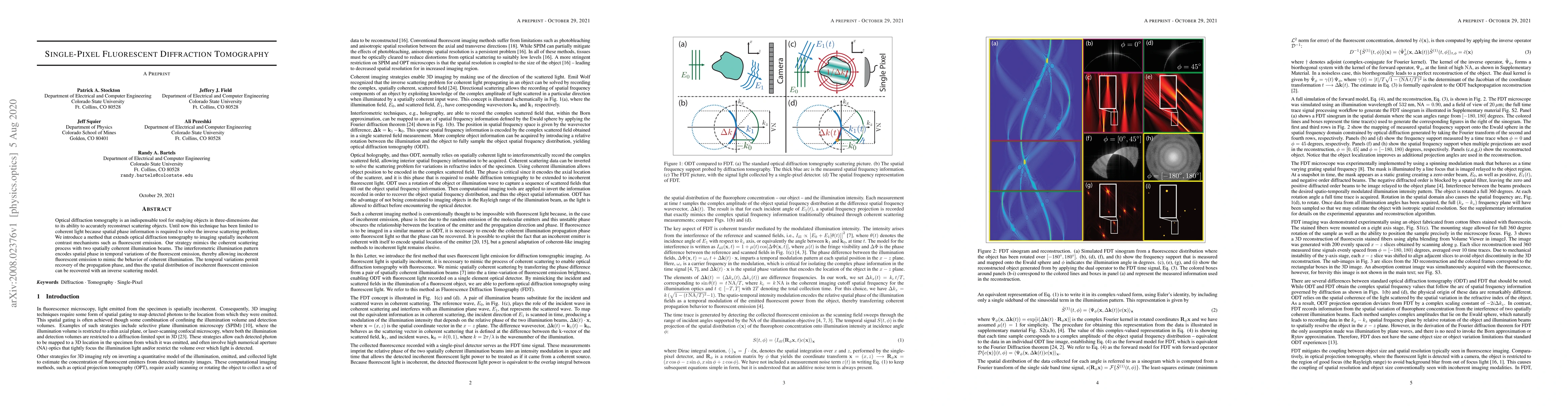
ArxivLens
ArXiv physics.optics eess.IV

Single-Pixel Fluorescent Diffraction Tomography

Ali Pezeshki, Jeff Squier, Jeffery J. Field, Patrick A. Stockton, Randy A. Bartels

2008.02376 Published Aug 7, 2020
View PDF

Publication

Published: Aug 07, 2020
Categories: physics.optics, eess.IV

Metrics

Source: ArXiv

AI Quick Summary

This paper introduces a novel method for single-pixel fluorescent diffraction tomography, enabling three-dimensional reconstruction of spatially incoherent fluorescent objects by mimicking coherent scattering with interferometric illumination patterns. This approach recovers the spatial distribution of fluorescent emission using temporal variations and an inverse scattering model.

Quick Answers

What is "Single-Pixel Fluorescent Diffraction Tomography" about?

This paper introduces a novel method for single-pixel fluorescent diffraction tomography, enabling three-dimensional reconstruction of spatially incoherent fluorescent objects by mimicking coherent scattering with interferometric illumination patterns. This approach recovers the spatial distribution of fluorescent emission using temporal variations and an inverse scattering model.

What methodology did the authors use?

The research introduces Single-Pixel Fluorescent Diffraction Tomography (FDT), a method that extends optical diffraction tomography to incoherent contrast mechanisms such as fluorescence and Raman scattering. It uses a continuous-wave laser, a spinning modulator disk, and spatial filtering to create a stationary reference beam and an angle-scanning beam, which mimic coherent scattering experiments. More in Methodology →

What are the key results?

3D reconstructions of fluorescent-stained cotton fibers were successfully demonstrated. — FDT works for both coherent and incoherent contrast mechanisms, including nonlinear mechanisms. More in Key Results →

Why is this work significant?

This research is significant as it extends the range of samples that can be imaged and provides richer information by co-registering multiple contrast distributions simultaneously, which is crucial for various applications in biological imaging and materials science. More in Significance →

What are the main limitations?

The experimental setup requires precise alignment and control of optical components. — The reconstruction algorithm's computational complexity may limit its practical implementation for large datasets. More in Limitations →

Paper Preview

Abstract

Optical diffraction tomography is an indispensable tool for studying objects in three-dimensions due to its ability to accurately reconstruct scattering objects. Until now this technique has been limited to coherent light because spatial phase information is required to solve the inverse scattering problem. We introduce a method that extends optical diffraction tomography to imaging spatially incoherent contrast mechanisms such as fluorescent emission. Our strategy mimics the coherent scattering process with two spatially coherent illumination beams. The interferometric illumination pattern encodes spatial phase in temporal variations of the fluorescent emission, thereby allowing incoherent fluorescent emission to mimic the behavior of coherent illumination. The temporal variations permit recovery of the propagation phase, and thus the spatial distribution of incoherent fluorescent emission can be recovered with an inverse scattering model.

Key Findings, in focus

Seven facets of this paper, analysed and brought into focus by AI.

AI-generated · verify against source Generated Sep 02, 2025
In focus · one-line synthesis

This research is significant as it extends the range of samples that can be imaged and provides richer information by co-registering multiple contrast distributions simultaneously, which is crucial for various applications in biological imaging and materials science.

01
MethodologyHow they did it

The research introduces Single-Pixel Fluorescent Diffraction Tomography (FDT), a method that extends optical diffraction tomography to incoherent contrast mechanisms such as fluorescence and Raman scattering. It uses a continuous-wave laser, a spinning modulator disk, and spatial filtering to create a stationary reference beam and an angle-scanning beam, which mimic coherent scattering experiments.

02
ResultsWhat they found
  • 3D reconstructions of fluorescent-stained cotton fibers were successfully demonstrated.
  • FDT works for both coherent and incoherent contrast mechanisms, including nonlinear mechanisms.
  • The technique decouples the need to reduce the numerical aperture of illumination as object size increases, expanding the range of samples that can be imaged.
  • FDT allows for rich information by co-registering multiple contrast distributions simultaneously.
03
SignificanceWhy it matters

This research is significant as it extends the range of samples that can be imaged and provides richer information by co-registering multiple contrast distributions simultaneously, which is crucial for various applications in biological imaging and materials science.

04
Technical ContributionsWhat's new technically

The main technical contribution is the development of a forward and inverse model for FDT, along with a novel experimental setup that encodes spatial phase information from incoherent fluorescent emission using interferometric illumination patterns.

05
NoveltyWhat sets it apart

The novelty of this work lies in its ability to apply optical diffraction tomography to incoherent contrast mechanisms like fluorescence, thereby expanding the applicability of this powerful imaging technique.

06
LimitationsWhat to be cautious about
  • The experimental setup requires precise alignment and control of optical components.
  • The reconstruction algorithm's computational complexity may limit its practical implementation for large datasets.
07
Future WorkWhere it goes next
  • Further optimization of the reconstruction algorithm for faster and more efficient processing.
  • Exploration of FDT's applicability to other incoherent contrast mechanisms beyond fluorescence and Raman scattering.

Impact

27
References

Paper Details

Paper ID: 2008.02376
License: http://arxiv.org/licenses/nonexclusive-distrib/1.0/
Categories:
physics.optics eess.IV

How to Cite This Paper

Reference manager: RIS EndNote

Key Terms

illumination (0.315)
coherent (0.275)
tomography (0.238)
emission (0.231)
scattering (0.231)
diffraction (0.229)
spatial (0.200)
spatially (0.199)

Citation Network

Current paper (gray), citations (green), references (blue)

Display is limited for performance on very large graphs.

Current Paper
Citations
References
Click to view

Related Papers

4 found


Discussion 0

This website uses cookies to ensure you get the best experience. Learn more