Summary
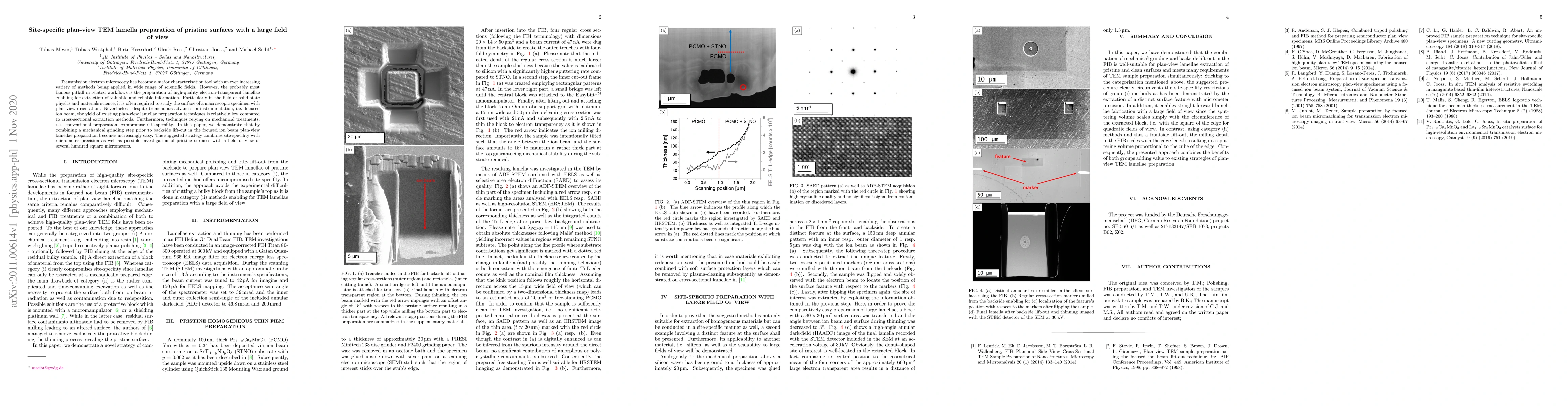
Transmission electron microscopy has become a major characterisation tool with an ever increasing variety of methods being applied in wide range of scientific fields. However, the probably most famous pitfall in related workflows is the preparation of high-quality electron-transparent lamellae enabling for extraction of valuable and reliable information. Particularly in the field of solid state physics and materials science, it is often required to study the surface of a macroscopic specimen with plan-view orientation. Nevertheless, despite tremendous advances in instrumentation, i.e. focused ion beam, the yield of existing plan-view lamellae preparation techniques is relatively low compared to cross-sectional extraction methods. Furthermore, techniques relying on mechanical treatments, i.e. conventional preparation, compromise site-specifity. In this paper, we demonstrate that by combining a mechanical grinding step prior to backside lift-out in the focused ion beam plan-view lamellae preparation becomes increasingly easy. The suggested strategy combines site-specifity with micrometer precision as well as possible investigation of pristine surfaces with a field of view of several hundred square micrometers.
AI Key Findings
Get AI-generated insights about this paper's methodology, results, and significance.
Paper Details
PDF Preview
Key Terms
Citation Network
Current paper (gray), citations (green), references (blue)
Display is limited for performance on very large graphs.
Similar Papers
Found 4 papersSite-Specific Plan-view (S)TEM Sample Preparation from Thin Films using a Dual-Beam FIB-SEM
Sreejith Nair, K. Andre Mkhoyan, Bharat Jalan et al.
An optimized TEM specimen preparation method of quantum nanostructures
Hongguang Wang, Jochen Mannhart, Peter A. van Aken et al.
No citations found for this paper.
Comments (0)