Slice Imputation: Intermediate Slice Interpolation for Anisotropic 3D Medical Image Segmentation
Publication
Metrics
AI Quick Summary
This paper proposes a novel frame-interpolation-based method for slice imputation to enhance segmentation accuracy in anisotropic 3D medical images by improving smoothness across all three axes. The method outperforms existing techniques by achieving better resolution and isotropic representations, leading to superior segmentation results in various medical imaging tasks.
Paper Preview
Abstract
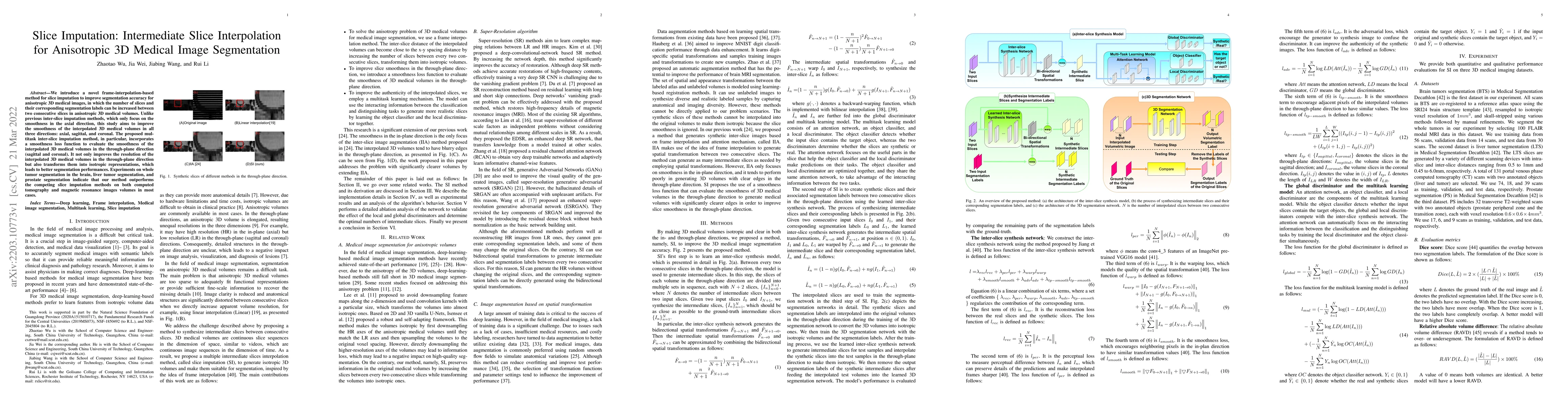
We introduce a novel frame-interpolation-based method for slice imputation to improve segmentation accuracy for anisotropic 3D medical images, in which the number of slices and their corresponding segmentation labels can be increased between two consecutive slices in anisotropic 3D medical volumes. Unlike previous inter-slice imputation methods, which only focus on the smoothness in the axial direction, this study aims to improve the smoothness of the interpolated 3D medical volumes in all three directions: axial, sagittal, and coronal. The proposed multitask inter-slice imputation method, in particular, incorporates a smoothness loss function to evaluate the smoothness of the interpolated 3D medical volumes in the through-plane direction (sagittal and coronal). It not only improves the resolution of the interpolated 3D medical volumes in the through-plane direction but also transforms them into isotropic representations, which leads to better segmentation performances. Experiments on whole tumor segmentation in the brain, liver tumor segmentation, and prostate segmentation indicate that our method outperforms the competing slice imputation methods on both computed tomography and magnetic resonance images volumes in most cases.
AI Key Findings
Get AI-generated insights about this paper's methodology, results, significance, and more — seven facets brought into focus.
Impact
Paper Details
Authors
PDF Preview
Key Terms
Citation Network
Current paper (gray), citations (green), references (blue)
Display is limited for performance on very large graphs.
Discussion 0