ArxivLens

Quick Summary:

This paper reports the first in vivo measurements of the longitudinal relaxation time T1 in human brain tissues using SQUID-based MRI at microtesla fields. The study reveals that T1 values are within 5% of the transverse relaxation time T2 at 46 microtesla, suggesting potential implications for imaging contrast in microtesla MRI.

Journal Reference: IEEE Trans. Appl. Supercond. 19:823-826, 2009
DOI: 10.1109/TASC.2009.2018764
arXiv Id: 0811.0389
Comments: To appear in Proceedings of 2008 Applied Superconductivity Conference
Date Published: 2009-07-18
Date Updated: 2009-07-18

Key Terms (TF-IDF):

mri (0.397)
relaxation (0.326)
brain (0.319)
relaxation (0.306)
brain (0.275)
values (0.247)
imaging (0.246)
values (0.221)
imaging (0.220)
measured (0.219)
living (0.204)
measured (0.201)
seven (0.196)
fields (0.196)
living (0.187)
magnetic (0.186)
field (0.182)
magnetic resonance (0.179)
longitudinal (0.176)
fields (0.176)
License: http://arxiv.org/licenses/nonexclusive-distrib/1.0/

Summary:

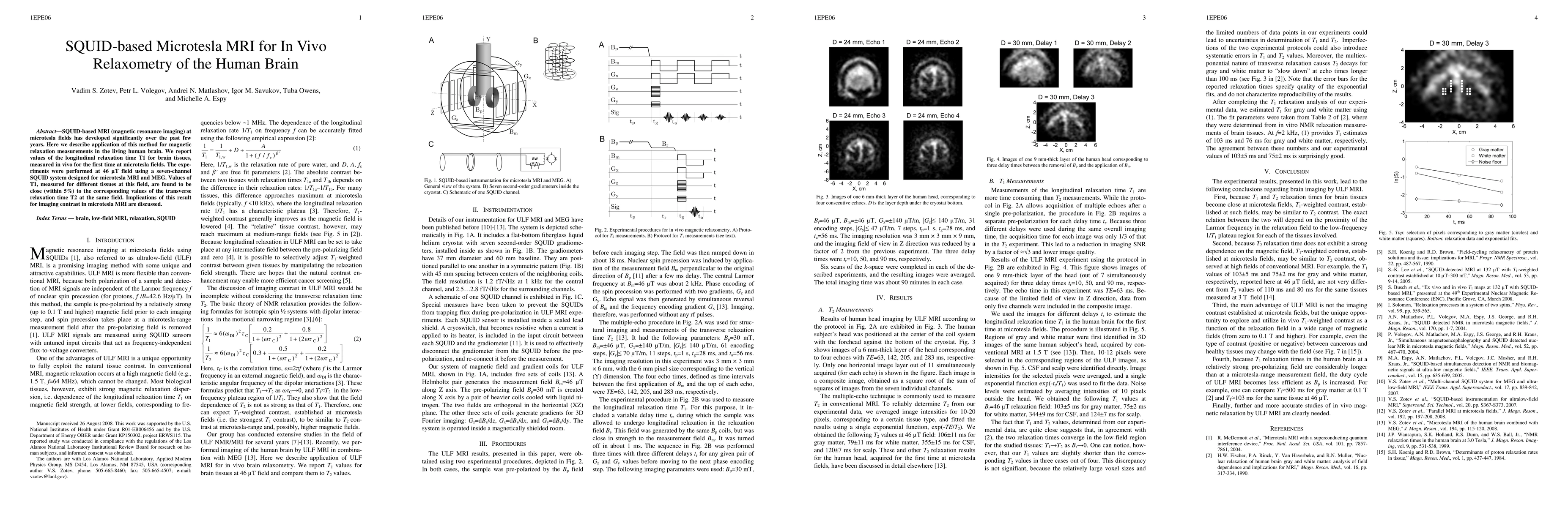
SQUID-based MRI (magnetic resonance imaging) at microtesla fields has developed significantly over the past few years. Here we describe application of this method for magnetic relaxation measurements in the living human brain. We report values of the longitudinal relaxation time T1 for brain tissues, measured in vivo for the first time at microtesla fields. The experiments were performed at 46 microtesla field using a seven-channel SQUID system designed for microtesla MRI and MEG. Values of T1, measured for different tissues at this field, are found to be close (within 5%) to the corresponding values of the transverse relaxation time T2 at the same field. Implications of this result for imaging contrast in microtesla MRI are discussed.

Citation & Reference Network Visualization

This graph shows how this paper is connected to other research. The current paper is shown in dark gray, papers that cite it are in green, and papers it references are in blue. You can:

Current Paper
Papers citing this work (Citations)
Papers cited by this work (References)

• Hover over any node to see the paper title

• Click a node to view that paper's details

• Drag nodes to rearrange the graph

• Use the zoom controls (bottom right) to zoom in/out

Citations and References

Title Authors Year Actions
Magnetic Resonance Imaging Using a Magnetoresistive Sensor With a Flux Transformer
D. Oyama, Y. Adachi, Naohiro Tsuyuguchi
2023
View Paper
Bringing MRI to low‐ and middle‐income countries: Directions, challenges and potential solutions
S. Murali, Hao Ding, Fope Adedeji, Cathy Qin, Johnes Obungoloch, I. Asllani, U. Anazodo, N. Ntusi, Regina Mammen, T. Niendorf, S. Adeleke
2023
View Paper
A Magnetic Resonance-Relaxometry-Based Technique to Identify Blood Products in Brain Parenchyma: An Experimental Study on a Rabbit Model
F. Del Signore, M. Vignoli, L. Della Salda, R. Tamburro, Andrea Paolini, I. Cerasoli, M. Chincarini, E. Rossi, N. Ferri, M. Romanucci, Ilaria Falerno, F. de Pasquale
2022
View Paper
Power Batteries Health Monitoring: A Magnetic Imaging Method Based on Magnetoelectric Sensors
Rui Chen, J. Jiao, Ziyun Chen, Yuhang Wang, Tingyu Deng, Wenning Di, Shunliang Zhu, Mingguang Gong, Li Lu, Xianyu Xie, Haosu Luo
2022
View Paper
Computational and Phantom-Based Feasibility Study of 3D dcNCI With Ultra-Low-Field MRI
N. Höfner, J. Storm, P. Hömmen, A. Cassarà, R. Körber
2021
View Paper
A numerical study of pre-polarisation switching in ultra-low field magnetic resonance imaging using dynamic permanent magnet arrays
Ruben Pellicer-Guridi, Michael W. Vogel, V. Vegh, Jiasheng Su, M. Rosen, D. Reutens
2020
View Paper
SQUID-Based Magnetic Resonance Imaging at Ultra-Low Field Using the Backprojection Method
Qingqian Guo, Chang-yu Ma, Xin Zhang, Yajie Xu, Meisheng Fan, P. Yu, Tao Hu, Yan Chang, Xiao-dong Yang
2020
View Paper
Low-Field MRI: How Low Can We Go? A Fresh View on an Old Debate
M. Sarracanie, N. Salameh
2020
View Paper
Evaluating the Performance of Ultra-Low-Field MRI for in-vivo 3D Current Density Imaging of the Human Head
P. Hömmen, A. Mäkinen, A. Hunold, R. Machts, J. Haueisen, K. Zevenhoven, R. Ilmoniemi, R. Körber
2020
View Paper
Towards direct neuronal current imaging via ultra-low-field MR
Nora Hofner, Rainer Korber, M. Burghoff
2019
View Paper
Demonstration of full tensor current density imaging using ultra-low field MRI.
P. Hömmen, J. Storm, N. Höfner, R. Körber
2019
View Paper
3D-Spatial encoding with permanent magnets for ultra-low field magnetic resonance imaging
Michael W. Vogel, Ruben Pellicer Guridi, Jiasheng Su, V. Vegh, D. Reutens
2019
View Paper
Feasibility of functional MRI at ultralow magnetic field via changes in cerebral blood volume
Kai Buckenmaier, A. F. Pedersen, P. SanGiorgio, K. Scheffler, J. Clarke, B. Inglis
2018
View Paper
Ultralow‐field and spin‐locking relaxation dispersion in postmortem pig brain
Hui Dong, Seong-Min Hwang, M. Wendland, Lixing You, J. Clarke, B. Inglis
2017
View Paper
Novel imaging using a MEG scanner, and MRI homogeneity improvement techniques
I. Vella
2017
View Paper
Spin Electronics for Biomagnetism and Nuclear Magnetic Resonance
myriam. lecoeur, R. Ayde, C. Fermon
2017
View Paper
Low-Cost High-Performance MRI
M. Sarracanie, Cris D. LaPierre, N. Salameh, D. Waddington, T. Witzel, M. Rosen
2015
View Paper
Signal Analysis and Liver Tumor Discrimination by Using a High-Tc SQUID-Based Low-Field NMR System in Hospital
S. Liao, J. Chieh, Yen-Ting Chou, Ming-Wei Wang, Hsin‐Hsien Chen, Kai-Wen Huang, Hong-Chang Yang, H. Horng
2015
View Paper
Progress Toward a Deployable SQUID-Based Ultra-Low Field MRI System for Anatomical Imaging
M. Espy, P. Magnelind, A. Matlashov, S. Newman, H. Sandin, L. Schultz, R. Sedillo, A. Urbaitis, P. Volegov
2015
View Paper
Prototype phantoms for characterization of ultralow field magnetic resonance imaging
M. Boss, J. Mates, Sarah E. Busch, P. SanGiorgio, S. Russek, Kai Buckenmaier, K. Irwin, Hsiao-mei Cho., G. Hilton, J. Clarke
2014
View Paper
Conductive shield for ultra-low-field magnetic resonance imaging: Theory and measurements of eddy currents.
K. Zevenhoven, Sarah E. Busch, M. Hatridge, Fredrik Oisjöen, R. Ilmoniemi, J. Clarke
2014
View Paper
Strong pulsed excitations using circularly polarized fields for ultra-low field NMR.
J. Shim, S. Lee, Kwon-Kyu Yu, Seong-Min Hwang, Kiwoong Kim
2014
View Paper
An advanced phantom study assessing the feasibility of neuronal current imaging by ultra-low-field NMR.
R. Körber, Jaakko O. Nieminen, N. Höfner, V. Jazbinsek, H. Scheer, Kiwoong Kim, M. Burghoff
2013
View Paper
MRI of the human brain at 130 microtesla
B. Inglis, Kai Buckenmaier, P. SanGiorgio, A. F. Pedersen, Matthew Nichols, J. Clarke
2013
View Paper
Temperature dependence of relaxation times and temperature mapping in ultra-low-field MRI.
P. Vesanen, K. Zevenhoven, Jaakko O. Nieminen, J. Dabek, L. Parkkonen, R. Ilmoniemi
2013
View Paper
Very low field magnetic resonance imaging with spintronic sensors.
Q. Herreros, H. Dyvorne, P. Campiglio, G. Jasmin-Lebras, A. Demonti, M. Pannetier-Lecoeur, C. Fermon
2013
View Paper
Magnetic-resonance imaging of the human brain with an atomic magnetometer.
I. Savukov, T. Karaulanov
2013
View Paper
Hybrid ultra‐low‐field MRI and magnetoencephalography system based on a commercial whole‐head neuromagnetometer
P. Vesanen, Jaakko O. Nieminen, K. Zevenhoven, J. Dabek, L. Parkkonen, A. Zhdanov, J. Luomahaara, J. Hassel, J. Penttilä, J. Simola, A. Ahonen, J. Mäkelä, R. Ilmoniemi
2013
View Paper
SQUID-detected ultra-low field MRI.
M. Espy, A. Matlashov, P. Volegov
2013
View Paper
WITHDRAWN: SQUID-detected ultra-low field MRI.
M. Espy, A. Matlashov, P. Volegov
2013
View Paper
Toward High Resolution Images With SQUID-Based Ultra-Low Field Magnetic Resonance Imaging
M. Espy, P. Magnelind, A. Matlashov, S. Newman, A. Urbaitis, P. Volegov
2013
View Paper
Magnetic resonance imaging at frequencies below 1 kHz.
I. Hilschenz, R. Körber, H. Scheer, T. Fedele, Hans-Helge Albrecht, Antonino Mario Cassará, S. Hartwig, L. Trahms, J. Haase, M. Burghoff
2013
View Paper
Gradient-excitation encoding combined with frequency and phase encodings for three-dimensional ultra-low-field MRI
J. Dabek, K. Zevenhoven, Jaakko O. Nieminen, P. Vesanen, R. Sepponen, R. Ilmoniemi
2012
View Paper
Current Awareness in NMR in Biomedicine
2010
View Paper
Microtesla MRI with dynamic nuclear polarization.
V. Zotev, T. Owens, A. Matlashov, I. Savukov, John J. Gomez, M. Espy
2009
View Paper
Neuronal Current Imaging with Ultralow-Field NMR Techniques
R. Körber, M. Burghoff, L. Trahms
2019
View Paper
Ultra-Low-Field MRI and Its Combination with MEG
L. Parkkonen, R. Ilmoniemi, F. Lin, M. Espy
2014
View Paper
SQUID-detected ultra-low field MRI.
M. Espy, A. Matlashov, P. Volegov
2013
View Paper
Combined ultra-low-field MRI and MEG: instrumentation and applications
P. Vesanen
2013
View Paper
Optical Atomic Magnetometry for Ultra-Low-Field NMR Detection
G. Bevilacqua, V. Biancalana, Y. Dancheva, L. Moi
2013
View Paper
The Spatial and Temporal Distortion of Magnetic Fields Applied Inside a Magnetically Shielded Room
P. Vesanen, Jaakko O. Nieminen, K. Zevenhoven, J. Dabek, J. Simola, J. Sarvas, R. Ilmoniemi
2012
View Paper
Ultra-low-field MRI: techniques and instrumentation for hybrid MEG-MRI
Jaakko O. Nieminen
2012
View Paper
Solving transient problems in ultra-low-field MRI
K. Zevenhoven
2011
View Paper
Magnetic Resonance Relaxometry at Low and Ultra low Fields.
P. Volegov, M. Flynn, R. Kraus, P. Magnelind, A. Matlashov, P. Nath, T. Owens, H. Sandin, I. Savukov, L. Schultz, A. Urbaitis, V. Zotev, M. Espy
2010
View Paper

Similar Papers

7 Tesla multimodal MRI dataset of ex-vivo human brain

Jing Zhang, Qinfeng Zhu, Haotian Li, Yao Shen, Sihui Li, Zhiyong Zhao, Dan Wu, Haoan Xu, Guojun Xu, Zuozhen Cao, Keqing Zhu

Published: 2024-12-06

Comments (0)

This website uses cookies to ensure you get the best experience. Learn more