Publication
Metrics
AI Quick Summary
This paper presents a cost-effective structured illumination microscopy (SIM) system using a Digital Micro-mirror Device (DMD) to rapidly switch high-frequency excitation patterns, achieving doubled spatial resolution. The proposed DMD-based laser interference SIM system (DMD-ISIM) successfully images nuclear pore complexes and microtubules in mammalian cells.
Paper Preview
Abstract
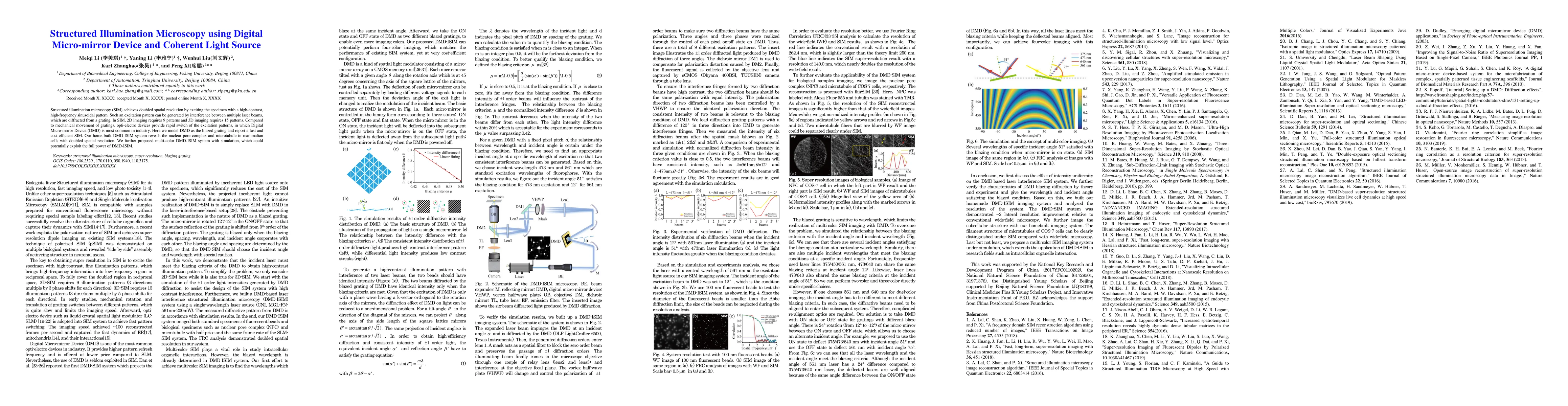
Structured illumination microscopy (SIM) achieves doubled spatial resolution by exciting the specimen with a high-contrast, high-frequency sinusoidal pattern. Such an excitation pattern can be generated by interference between multiple laser beams, which are diffracted from a grating. In SIM, 2D imaging requires 9 patterns and 3D imaging requires 15 patterns. Compared to mechanical movement of gratings, opti-electro devices provide rapid switch of the excitation patterns, in which Digital Micro-mirror Device (DMD) is most common in industry. Here we model DMD as the blazed grating and report a fast and cost-efficient SIM. Our home-built DMD-based laser interference structured illumination microscopy (DMD-ISIM) system reveals the nuclear pore complex and microtubule in mammalian cells with doubled spatial resolution. We further proposed multi-color DMD-ISIM system with simulation, which could potentially exploit the full power of DMD-ISIM.
AI Key Findings
Get AI-generated insights about this paper's methodology, results, significance, and more — seven facets brought into focus.
Impact
Paper Details
Authors
PDF Preview
Key Terms
Citation Network
Current paper (gray), citations (green), references (blue)
Display is limited for performance on very large graphs.
Discussion 0