Structured illumination microscopy with extended axial resolution through mirrored illumination
Publication
Metrics
AI Quick Summary
This paper proposes a dual-objective scheme for structured illumination microscopy that enhances axial resolution without the complexity of interferometric methods, achieving resolutions better than 125 nm while maintaining the speed of wide-field fluorescence microscopy.
Paper Preview
Abstract
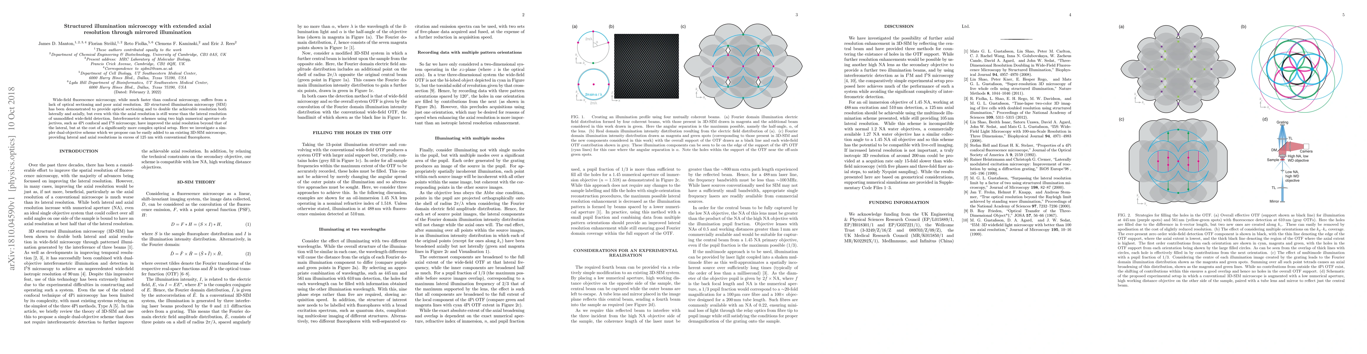
Wide-field fluorescence microscopy, while much faster than confocal microscopy, suffers from a lack of optical sectioning and poor axial resolution. 3D structured illumination microscopy (SIM) has been demonstrated to provide optical sectioning and to double the achievable resolution both laterally and axially, but even with this the axial resolution is still worse than the lateral resolution of unmodified wide-field detection. Interferometric schemes using two high numerical aperture objectives, such as 4Pi confocal and I5S microscopy, have improved the axial resolution beyond that of the lateral, but at the cost of a significantly more complex optical setup. Here we investigate a simpler dual-objective scheme which we propose can be easily added to an existing 3D-SIM microscope, providing lateral and axial resolutions in excess of 125 nm with conventional fluorophores.
AI Key Findings
Get AI-generated insights about this paper's methodology, results, significance, and more — seven facets brought into focus.
Impact
Paper Details
PDF Preview
Key Terms
Citation Network
Current paper (gray), citations (green), references (blue)
Display is limited for performance on very large graphs.
Discussion 0