Sub-nanometer depth resolution and single dopant visualization achieved by tilt-coupled multislice electron ptychography
Publication
Metrics
AI Quick Summary
This research presents a novel algorithm for multislice electron ptychography that enhances depth resolution to sub-nanometer scales using minimal tilt angles and few projections, significantly improving single dopant visualization in materials science. The method successfully detects dilute praseodymium dopants in Ca2Co2O5 and reveals lattice distortions.
Paper Preview
Abstract
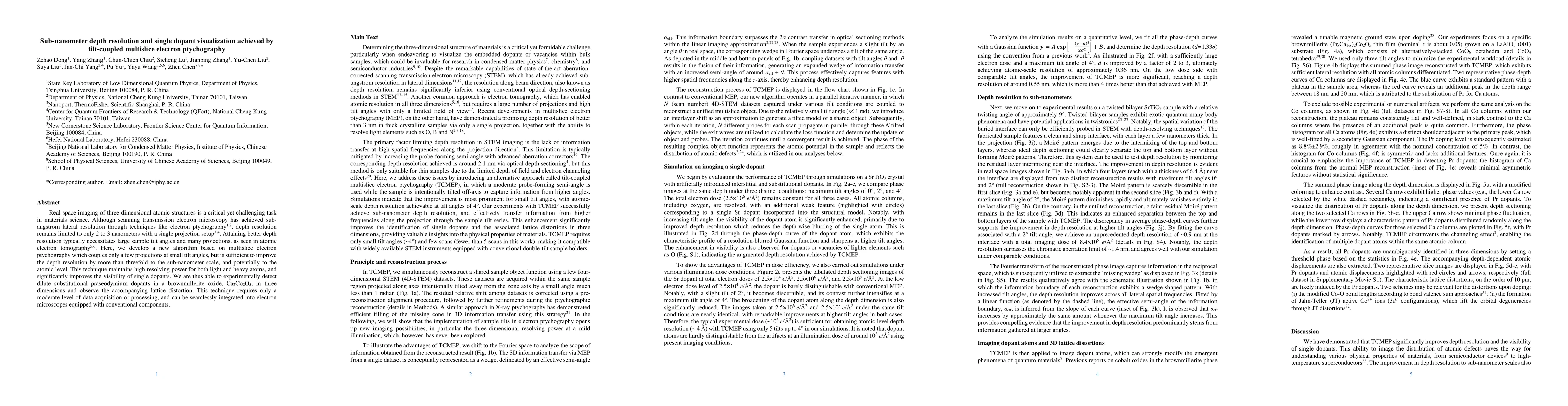
Real-space imaging of three-dimensional atomic structures is a critical yet challenging task in materials science. Although scanning transmission electron microscopy has achieved sub-angstrom lateral resolution through techniques like electron ptychography1,2, depth resolution remains limited to only 2 to 3 nanometers with a single projection setup3,4. Attaining better depth resolution typically necessitates large sample tilt angles and many projections, as seen in atomic electron tomography5,6. Here, we develop a new algorithm based on multislice electron ptychography which couples only a few projections at small tilt angles, but is sufficient to improve the depth resolution by more than threefold to the sub-nanometer scale, and potentially to the atomic level. This technique maintains high resolving power for both light and heavy atoms, and significantly improves the visibility of single dopants. We are thus able to experimentally detect dilute substitutional praseodymium dopants in a brownmillerite oxide, Ca2Co2O5, in three dimensions and observe the accompanying lattice distortion. This technique requires only a moderate level of data acquisition or processing, and can be seamlessly integrated into electron microscopes equipped with conventional components.
AI Key Findings
Get AI-generated insights about this paper's methodology, results, significance, and more — seven facets brought into focus.
Impact
Paper Details
Authors
PDF Preview
Key Terms
Citation Network
Current paper (gray), citations (green), references (blue)
Display is limited for performance on very large graphs.
Discussion 0