Quick Summary:

This research introduces "super phantoms" for advanced testing of medical imaging technologies, surpassing standard phantoms by replicating complex tissue properties. These advanced models, including computer models and ex-vivo organs, facilitate iterative improvements and innovation before in-vivo studies, supported by centralized facilities for multiple institutions.

Summary

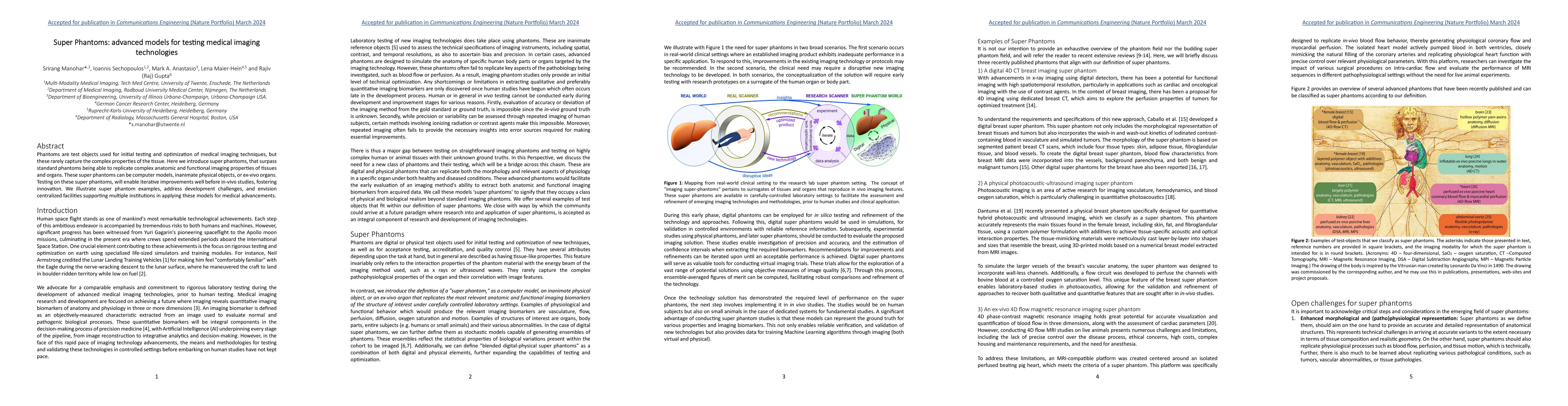
Phantoms are test objects used for initial testing and optimization of medical imaging techniques, but these rarely capture the complex properties of the tissue. Here we introduce super phantoms, that surpass standard phantoms being able to replicate complex anatomic and functional imaging properties of tissues and organs. These super phantoms can be computer models, inanimate physical objects, or ex-vivo organs. Testing on these super phantoms, will enable iterative improvements well before in-vivo studies, fostering innovation. We illustrate super phantom examples, address development challenges, and envision centralized facilities supporting multiple institutions in applying these models for medical advancements.

AI Key Findings

Get AI-generated insights about this paper's methodology, results, and significance.

Paper Details

PDF Preview

Download PDF

Key Terms

super (0.374)
super (0.374)
medical (0.295)
medical (0.280)
testing (0.272)
testing (0.257)
imaging (0.244)
imaging (0.234)
objects (0.223)
objects (0.220)
advancements (0.201)
facilities (0.201)
tissue (0.195)
centralized (0.190)
complex (0.188)
advancements (0.184)
supporting (0.184)
complex (0.183)
supporting (0.181)
iterative (0.169)

Citation Network

Current paper (gray), citations (green), references (blue)

Current Paper
Citations
References
Click to view
Title Authors Year Actions
Anthropomorphic Head MRI Phantoms: Technical Development, Brain Imaging Applications, and Future Prospects.
Lijun Deng, Ting Wu, Fei Wu, Lang Xiong, Hui Yang, Qingping Chen, Yupeng Liao
2025
View Paper
PixelPrint4D: A 3D Printing Method of Fabricating Patient-Specific Deformable CT Phantoms for Respiratory Motion Applications.
Jessica Y Im, Neghemi Micah, A. Perkins, Kai Mei, Michael Geagan, Leonid Roshkovan, Peter B. Noël
2025
View Paper
Investigating 4D respiratory cone-beam CT imaging for thoracic interventions on robotic C-arm systems: a deformable phantom study
Tess Reynolds, Owen Dillon, Yiqun Q Ma, Nicholas Hindley, J. Stayman, M. Bazalova-Carter
2024
View Paper
High-fidelity anatomical phantoms for MRI practical training.
Y. Habeeb, P. Zorn, F. Blindauer, N. Kharouf, D. Semeril, G. Bierry, S. Kremer, J. Dillenseger
2024
View Paper
Tailored 3D microphantoms: An essential tool for quantitative phase tomography analysis of organoids
M. Ziemczonok, S. Desissaire, Jeremy Neri, A. Kuś, L. Hervé, C. Fiche, G. Godefroy, Marie Fackeure, Damien Sery, W. Krauze, Kiran Padmanabhan, C. Paviolo, Małgorzata Kujawińska
2024
View Paper
TEMPy: a toolkit for the modeling of weighted tissue equivalent material in diagnostic imaging
Gisell R Boiset, Raphael Moratta, Elisabeth M Yoshimura, P. R. Costa
2024
View Paper

Comments (0)

This website uses cookies to ensure you get the best experience. Learn more