Publication

Published: Feb 20, 2026
Categories: physics.optics

Metrics

Source: ArXiv

Quick Actions

View PDF

Quick Answers

What methodology did the authors use?

Structured illumination X‑ray microscopy using a stepped 2D binary phase grating and Fourier-domain decomposition to recover high‑frequency sample replicas, followed by a generalized Wiener filter reconstruction and phase‑optimized separation matrix. More in Methodology →

What are the key results?

Achieved a 2.1× resolution gain on a test pattern, extending the effective pass‑band to 0.64 µm pixel size. — Demonstrated seamless integration of the technique into standard X‑ray tomography, yielding super‑resolved volumetric data with a 2.04 µm effective pixel size. More in Key Results →

Why is this work significant?

The work removes detector pixel‑size limits for photon‑counting X‑ray detectors, enabling non‑destructive testing and high‑resolution biomedical imaging without increasing magnification or exposure. More in Significance →

What are the main limitations?

Requires a high‑visibility, precisely aligned Talbot‑based grating, limiting applicability to well‑controlled beamlines. — Acquisition demands multiple images per illumination step, increasing data volume and potential radiation dose. More in Limitations →

Paper Preview

Abstract

We present a structured-illumination technique for full-field super-resolution transmission X-ray microscopy, which employs Fourier spectral decomposition inspired by established methods in visible-light microscopy. A 2D grating creating this illumination is stepped across one period to acquire a set of images at unique illumination positions. The Fourier domain of each image is described as a linear combination of replicated sample information at each frequency harmonic. As this superposition is created independently of detection, it contains spatial information exceeding native detector resolution. Recovering the encoded high-frequency components enables the population of an expanded frequency space. We demonstrate the presence of additional sample information in the Fourier spectrum and introduce a method to recover it. We achieve a resolution improvement by a factor of 2.1 for the projection image of a resolution test pattern. We further demonstrate seamless integration into standard X-ray tomography acquisition schemes. The acquisition is inherently multimodal, as phase-contrast and dark-field images can be computed from the same data using methods such as unified modulated pattern analysis, while providing an additional super-resolved transmission channel. These results indicate broad potential for non-destructive testing and biomedical imaging, as they alleviate pixel-size limitations in photon-counting detectors and sample-size restrictions imposed by optical magnification.
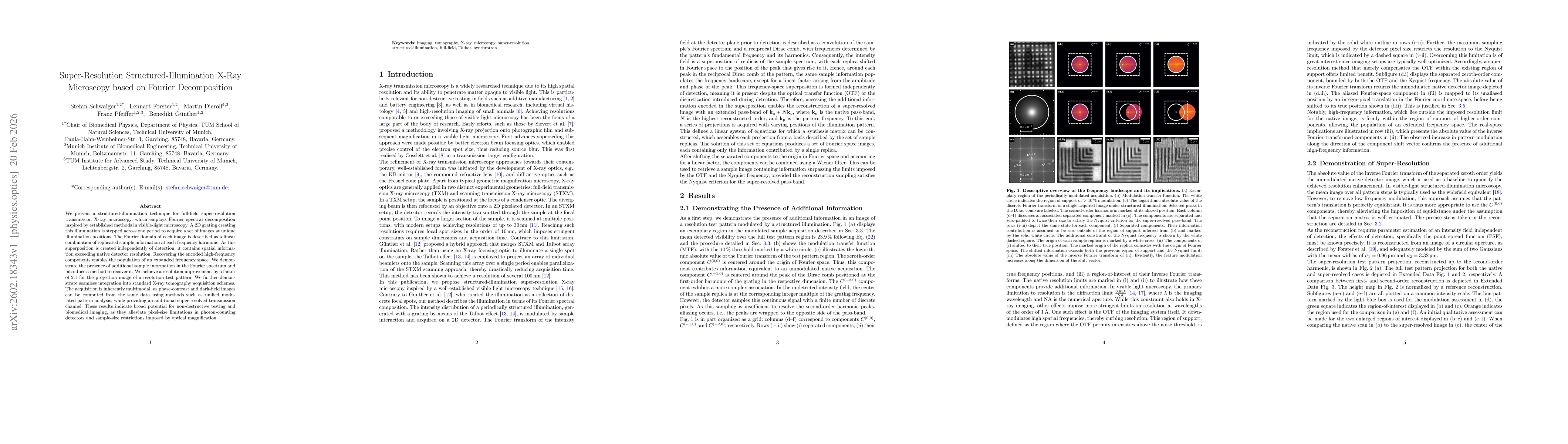
AI Key Findings

Generated Feb 24, 2026

Methodology — What approach did the authors take?

Structured illumination X‑ray microscopy using a stepped 2D binary phase grating and Fourier-domain decomposition to recover high‑frequency sample replicas, followed by a generalized Wiener filter reconstruction and phase‑optimized separation matrix.

Key Results — What are the main findings?

  • Achieved a 2.1× resolution gain on a test pattern, extending the effective pass‑band to 0.64 µm pixel size.
  • Demonstrated seamless integration of the technique into standard X‑ray tomography, yielding super‑resolved volumetric data with a 2.04 µm effective pixel size.
  • Provided multimodal imaging (transmission, phase‑contrast, dark‑field) from the same dataset, enabling richer sample characterization.
  • Validated the method on both laboratory and synchrotron beamlines, confirming robustness across different photon energies and detector configurations.

Significance — Why does this research matter?

The work removes detector pixel‑size limits for photon‑counting X‑ray detectors, enabling non‑destructive testing and high‑resolution biomedical imaging without increasing magnification or exposure.

Technical Contribution — What is the technical contribution?

A Fourier‑domain framework that models structured illumination as a linear superposition of shifted sample spectra, enabling explicit recovery of high‑frequency components via a phase‑optimized separation matrix and generalized Wiener filtering.

Novelty — What is new about this work?

First demonstration of full‑field, super‑resolved X‑ray transmission microscopy using structured illumination and Fourier decomposition, with simultaneous phase‑contrast and dark‑field imaging and direct compatibility with conventional tomography workflows.

Limitations — What are the limitations of this study?

  • Requires a high‑visibility, precisely aligned Talbot‑based grating, limiting applicability to well‑controlled beamlines.
  • Acquisition demands multiple images per illumination step, increasing data volume and potential radiation dose.
  • Phase‑optimization accuracy is critical; misestimation can leave residual grating artifacts and reduce resolution.
  • Aliasing from marginally unresolved higher harmonics can degrade reconstruction if not properly corrected.

Future Work — What did the authors propose for future work?

  • Extend the method to higher‑order harmonics and adaptive grating periods for even greater resolution gains.
  • Develop faster, single‑shot or compressed‑sensing acquisition schemes to reduce dose and acquisition time.
  • Integrate the technique with other contrast mechanisms (e.g., X‑ray fluorescence) for multimodal 3D imaging.
  • Automate phase‑estimation and alignment procedures to make the system more user‑friendly for routine industrial use.

Paper Details

Paper ID: 2602.18343
Categories:
physics.optics

How to Cite This Paper

BibTeX
@article{günther2026super,
  title         = {Super-Resolution Structured-Illumination X-Ray Microscopy based on Fourier Decomposition},
  author        = {Günther, Benedikt and Pfeiffer, Franz and Forster, Lennart and others},
  year          = {2026},
  eprint        = {2602.18343},
  archivePrefix = {arXiv},
  primaryClass  = {physics.optics},
}
APA 7
Günther, B., Pfeiffer, F., Forster, L., Dierolf, M., & Schwaiger, S. (2026). Super-Resolution Structured-Illumination X-Ray Microscopy based on Fourier Decomposition. arXiv. https://arxiv.org/abs/2602.18343
MLA 9
Günther, Benedikt, et al. "Super-Resolution Structured-Illumination X-Ray Microscopy based on Fourier Decomposition." arXiv, 2026, arxiv.org/abs/2602.18343.

Similar Papers

Found 4 papers

Comments (0)

This website uses cookies to ensure you get the best experience. Learn more