Academic Profile
Statistics
Similar Authors
Papers on arXiv
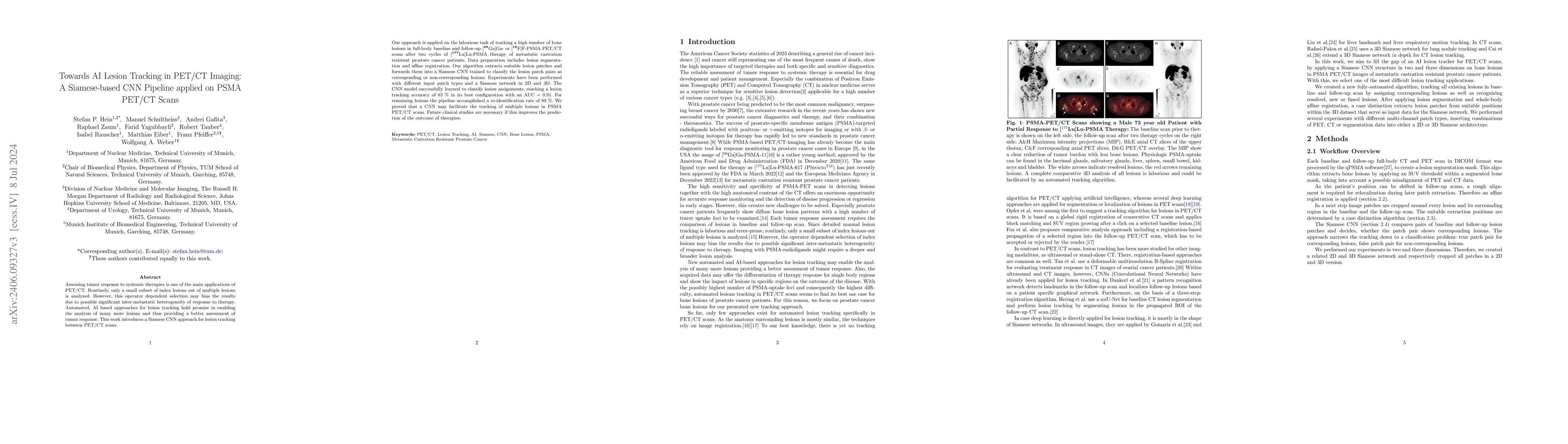
Assessing tumor response to systemic therapies is one of the main applications of PET/CT. Routinely, only a small subset of index lesions out of multiple lesions is analyzed. However, this operator de...
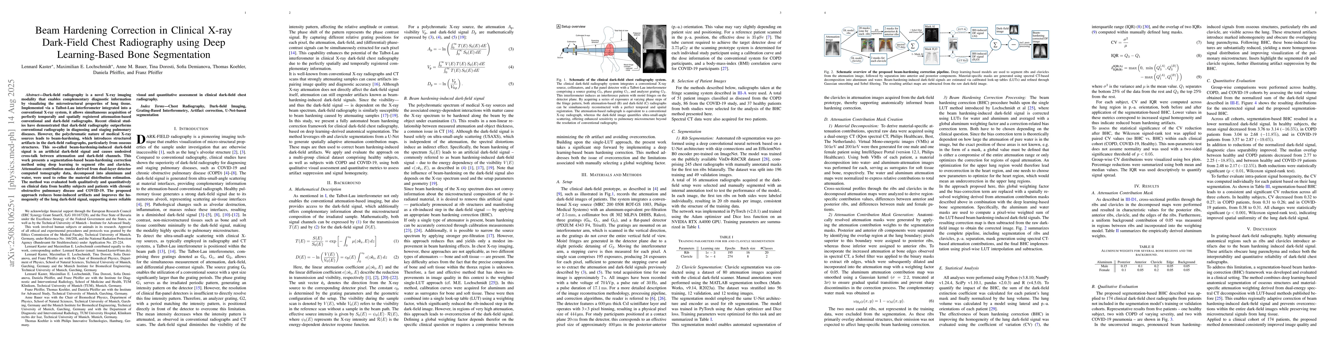
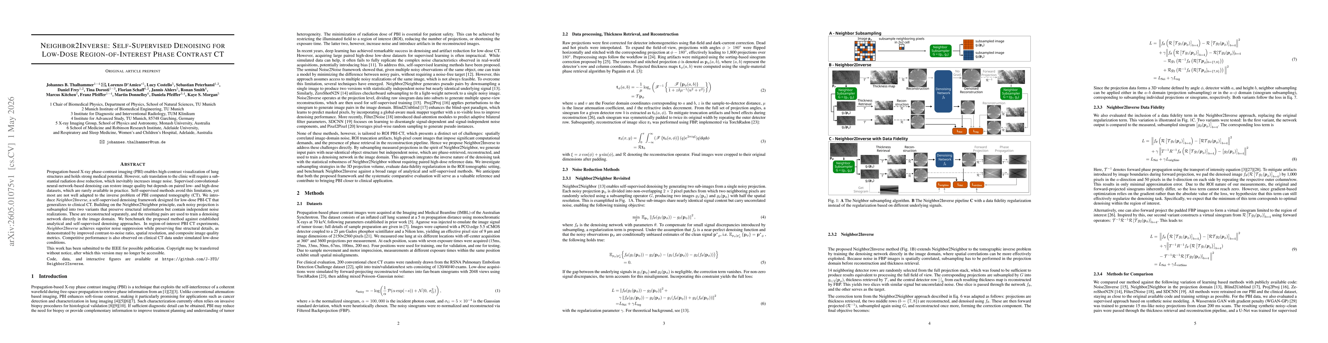
Background: We aimed at improving image quality (IQ) of sparse-view computed tomography (CT) images using a U-Net for lung metastasis detection and determining the best tradeoff between number of vi...
Tomographic imaging systems are expected to work with a wide range of samples that house complex structures and challenging material compositions, which can influence image quality in a bad way. Com...
This work presents methods for the seamless execution of arbitrary spherical trajectories with a seven-degree-of-freedom robotic arm as a sample holder. The sample holder is integrated into an exist...
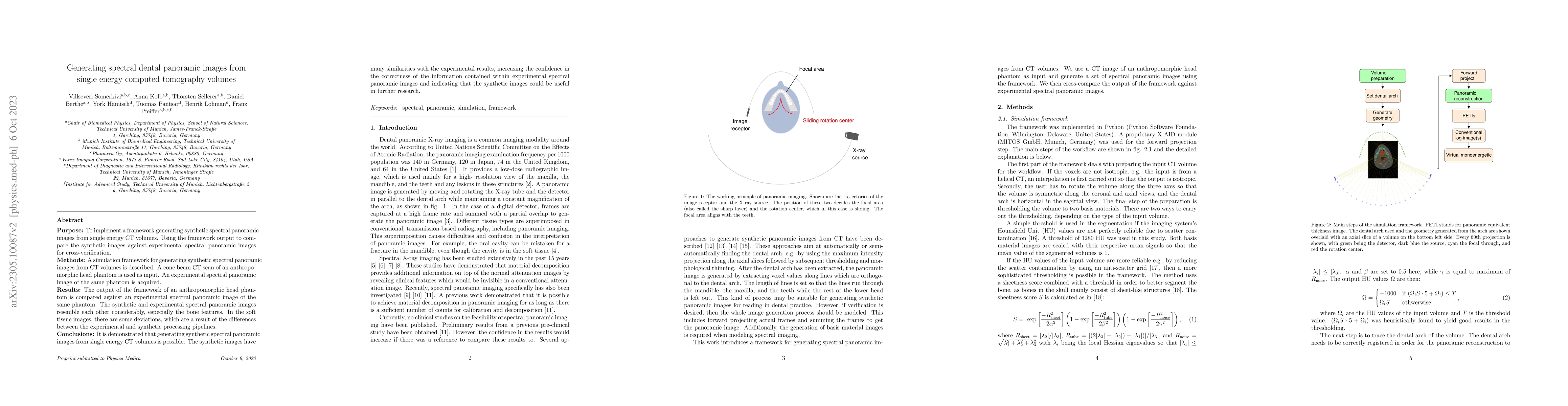
Purpose: To implement a framework generating synthetic spectral panoramic images from single energy CT volumes. Using the framework output to compare the synthetic images against experimental spectr...
Purpose: Sparse-view computed tomography (CT) is an effective way to reduce dose by lowering the total number of views acquired, albeit at the expense of image quality, which, in turn, can impact th...
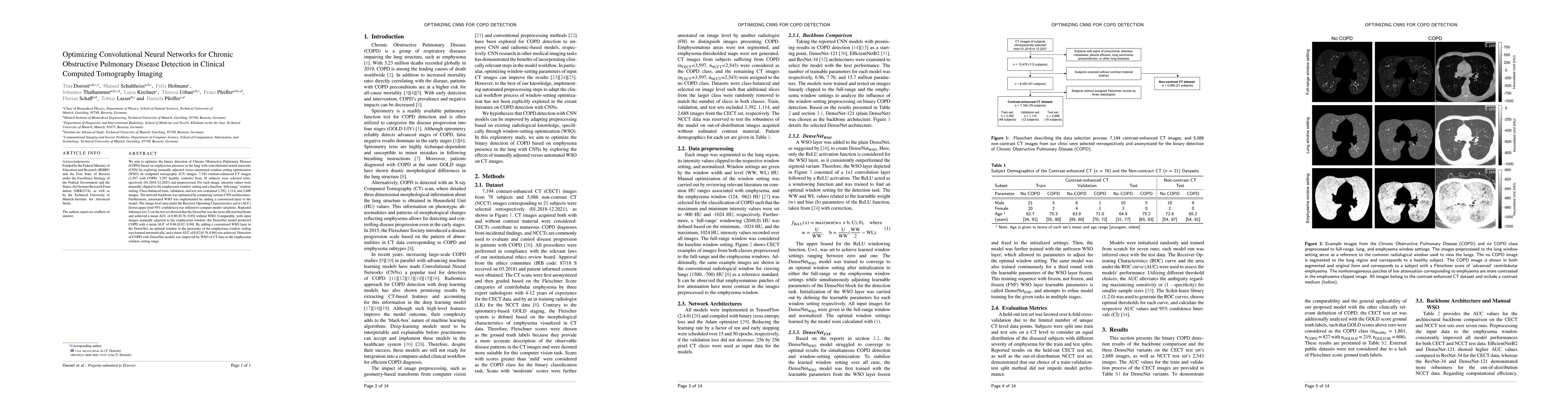
We aim to optimize the binary detection of Chronic Obstructive Pulmonary Disease (COPD) based on emphysema presence in the lung with convolutional neural networks (CNN) by exploring manually adjuste...
Deep learning based solutions are being succesfully implemented for a wide variety of applications. Most notably, clinical use-cases have gained an increased interest and have been the main driver b...
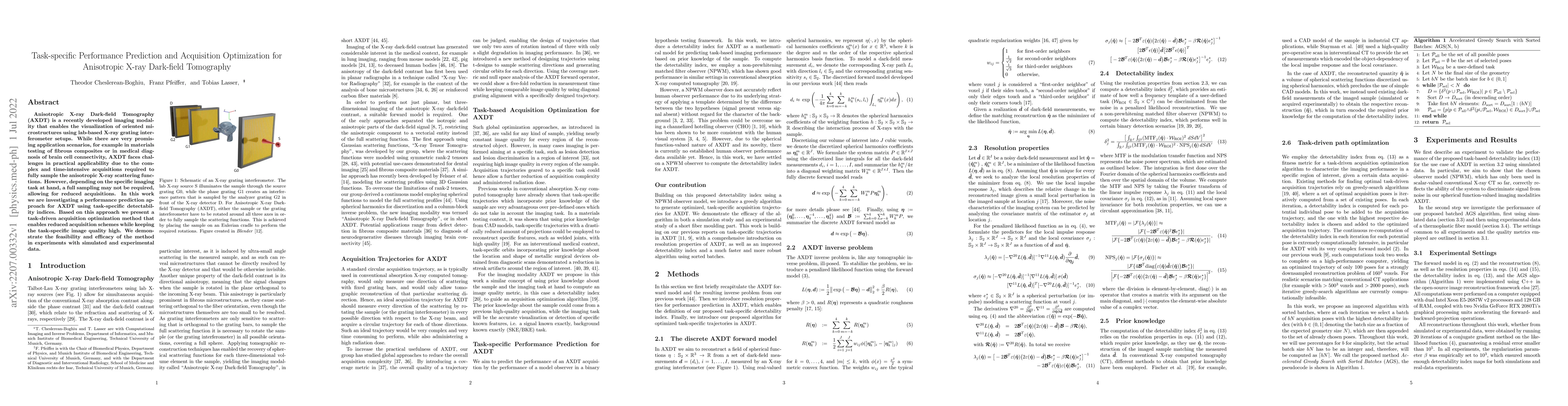
Anisotropic X-ray Dark-field Tomography (AXDT) is a recently developed imaging modality that enables the visualization of oriented microstructures using lab-based X-ray grating interferometer setups...
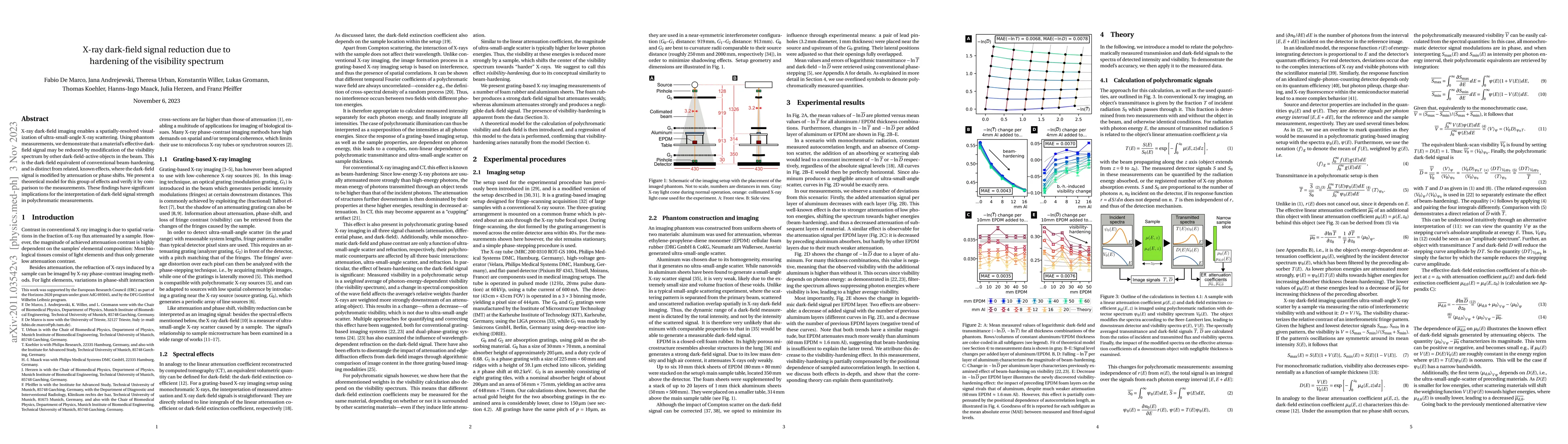
X-ray dark-field imaging enables a spatially-resolved visualization of ultra-small-angle X-ray scattering. Using phantom measurements, we demonstrate that a material's effective dark-field signal ma...
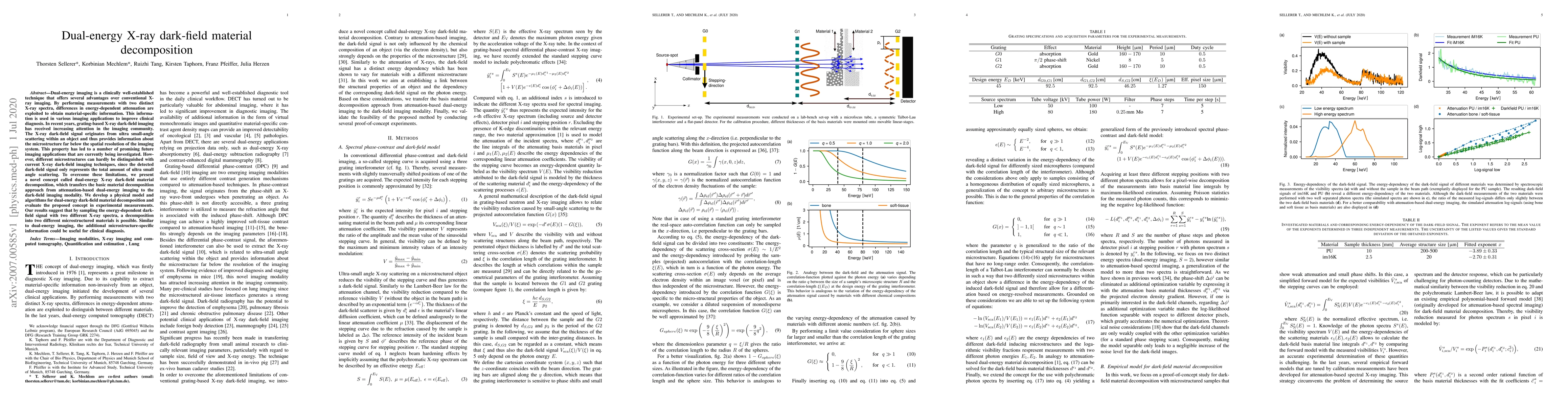
Dual-energy imaging is a clinically well-established technique that offers several advantages over conventional X-ray imaging. By performing measurements with two distinct X-ray spectra, differences...
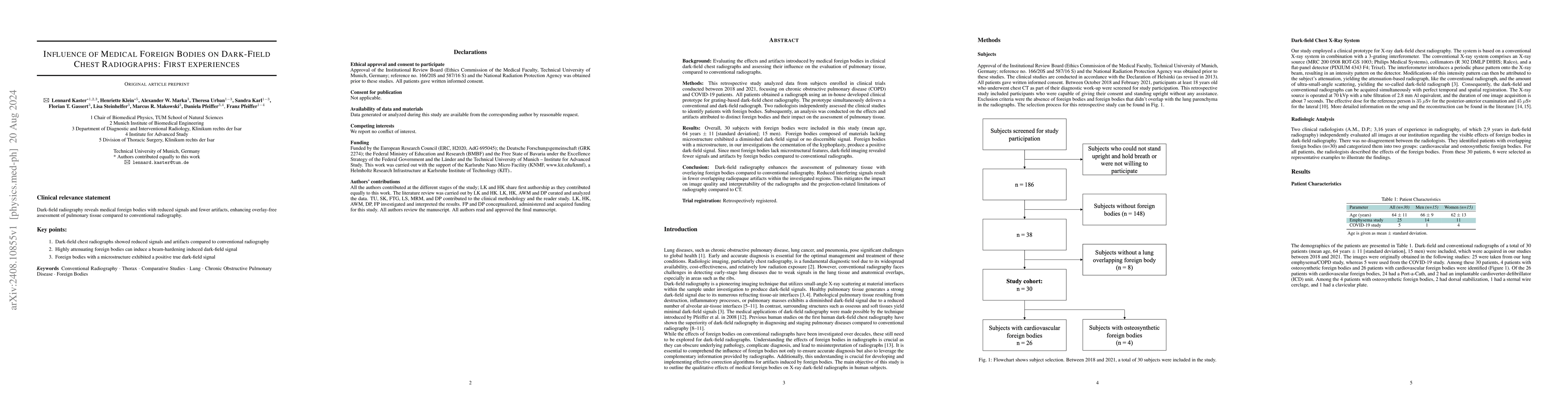
Objectives: Evaluating the effects and artifacts introduced by medical foreign bodies in clinical dark-field chest radiographs and assessing their influence on the evaluation of pulmonary tissue, comp...
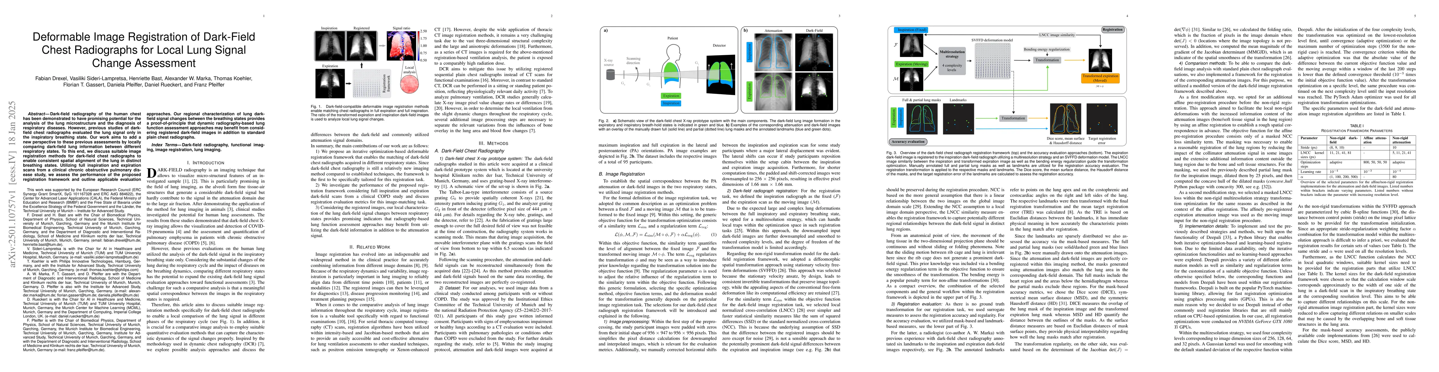
Dark-field radiography of the human chest has been demonstrated to have promising potential for the analysis of the lung microstructure and the diagnosis of respiratory diseases. However, previous stu...
Background: Material structures at the micrometer scale cause ultra-small-angle X-ray scattering, e.g., seen in lung tissue or plastic foams. In grating-based X-ray imaging, this causes a reduction of...
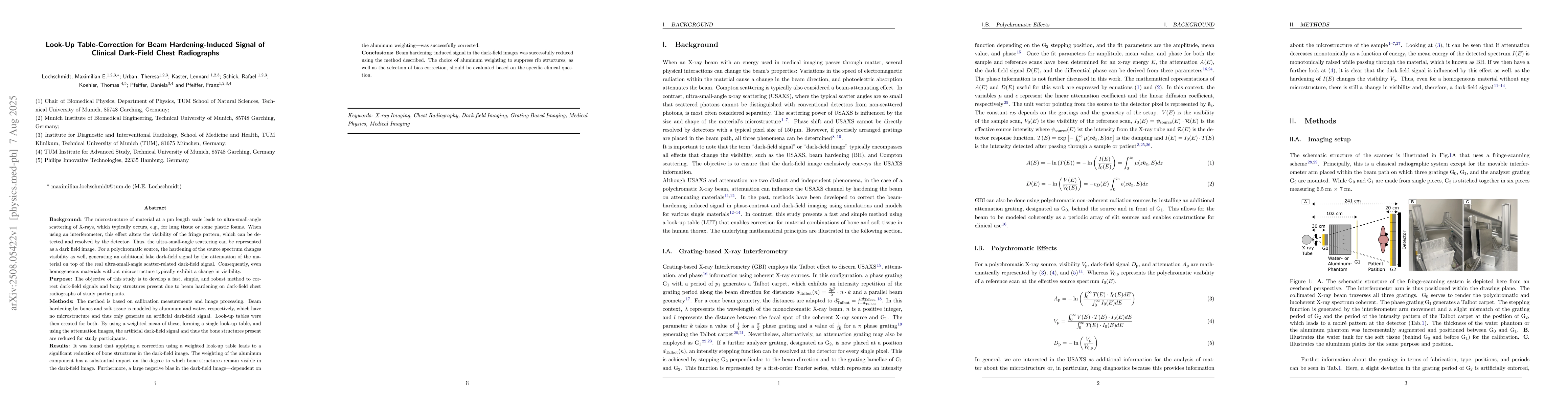
Dark-field radiography is a novel X-ray imaging modality that enables complementary diagnostic information by visualizing the microstructural properties of lung tissue. Implemented via a Talbot-Lau in...
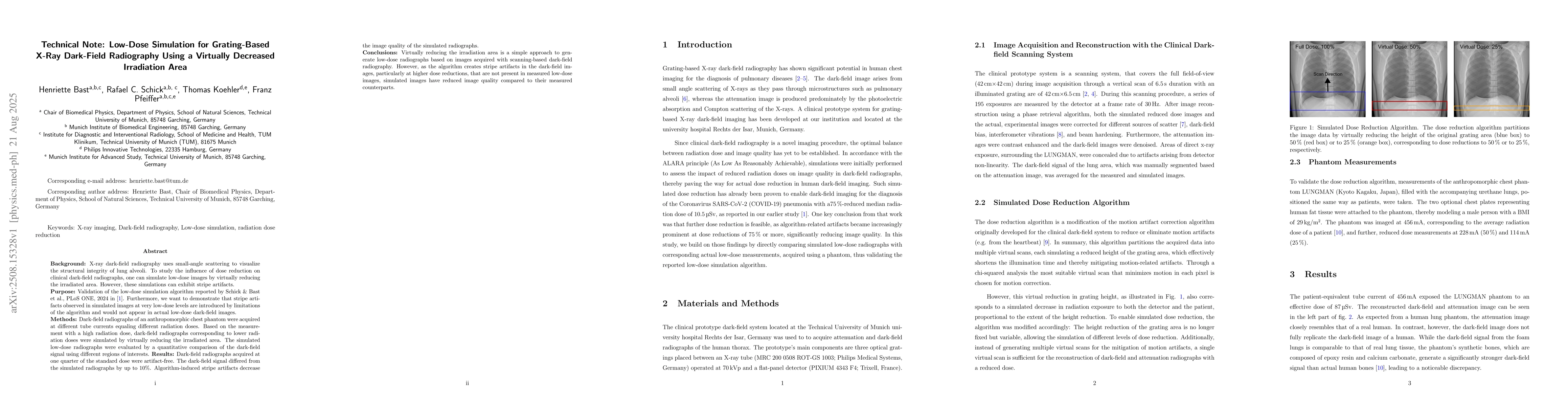
Background: X-ray dark-field radiography uses small-angle scattering to visualize the structural integrity of lung alveoli. To study the influence of dose reduction on clinical dark-field radiographs,...
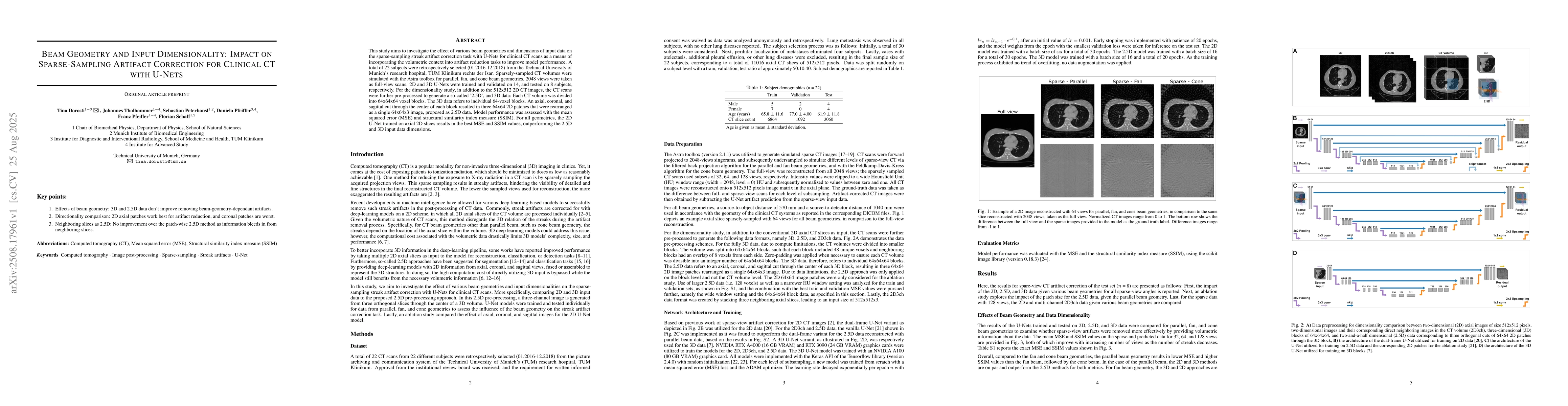
This study aims to investigate the effect of various beam geometries and dimensions of input data on the sparse-sampling streak artifact correction task with U-Nets for clinical CT scans as a means of...
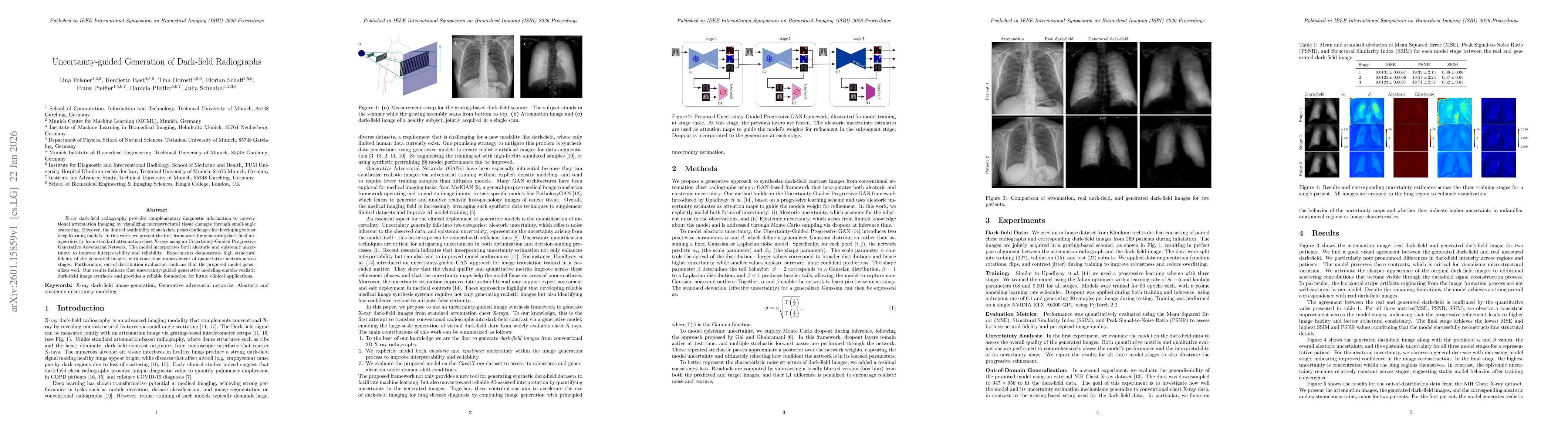
X-ray dark-field radiography provides complementary diagnostic information to conventional attenuation imaging by visualizing microstructural tissue changes through small-angle scattering. However, th...
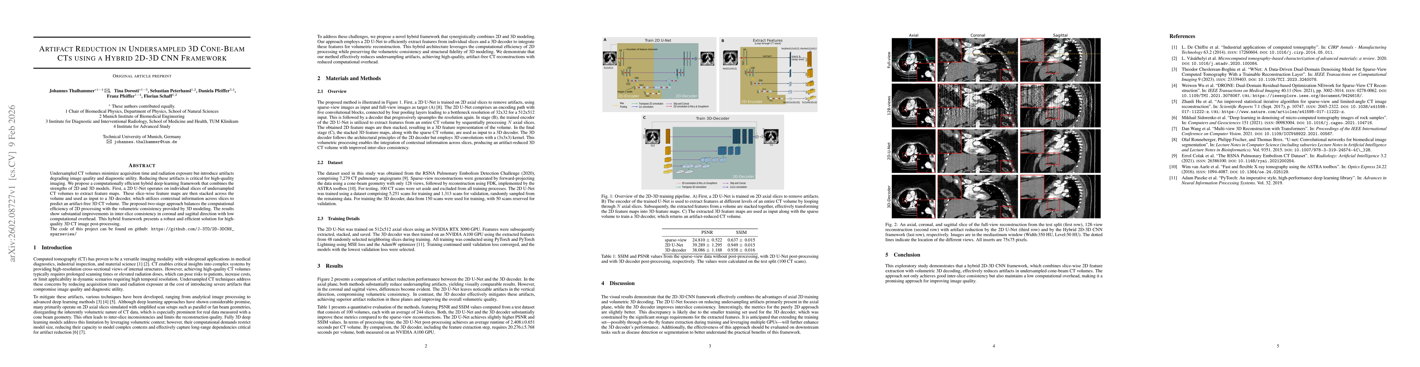
Undersampled CT volumes minimize acquisition time and radiation exposure but introduce artifacts degrading image quality and diagnostic utility. Reducing these artifacts is critical for high-quality i...
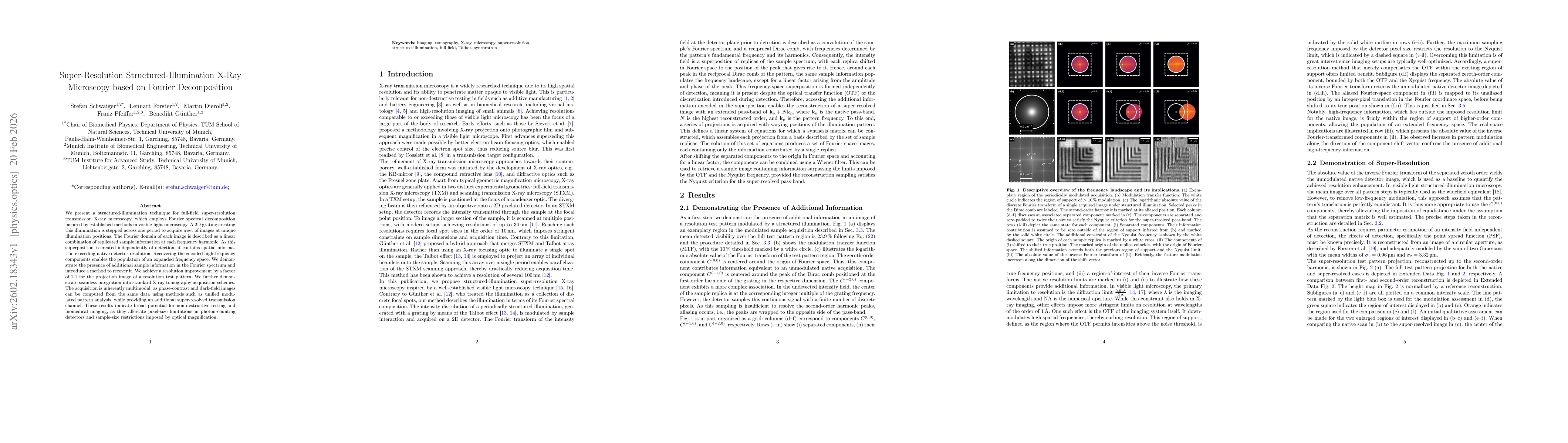
We present a structured-illumination technique for full-field super-resolution transmission X-ray microscopy, which employs Fourier spectral decomposition inspired by established methods in visible-li...
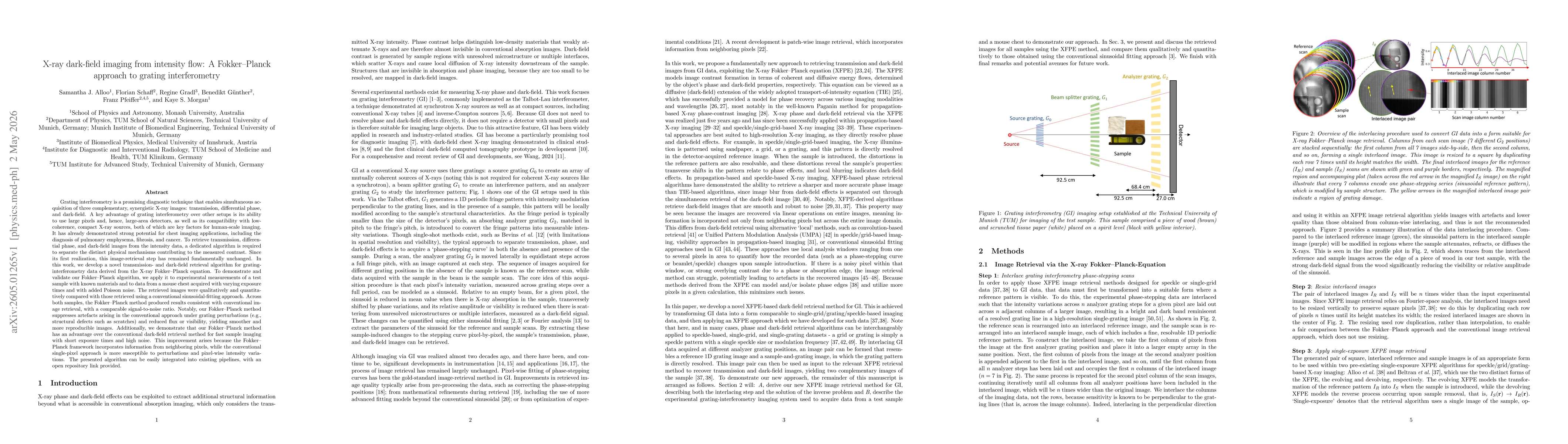
Grating interferometry is a promising diagnostic technique that enables simultaneous acquisition of three complementary, synergistic X-ray images: transmission, differential phase, and dark-field. Its...
Propagation-based X-ray phase-contrast imaging (PBI) enables high-contrast visualization of lung structures and holds strong medical potential. However, safe translation to the clinic will require a s...
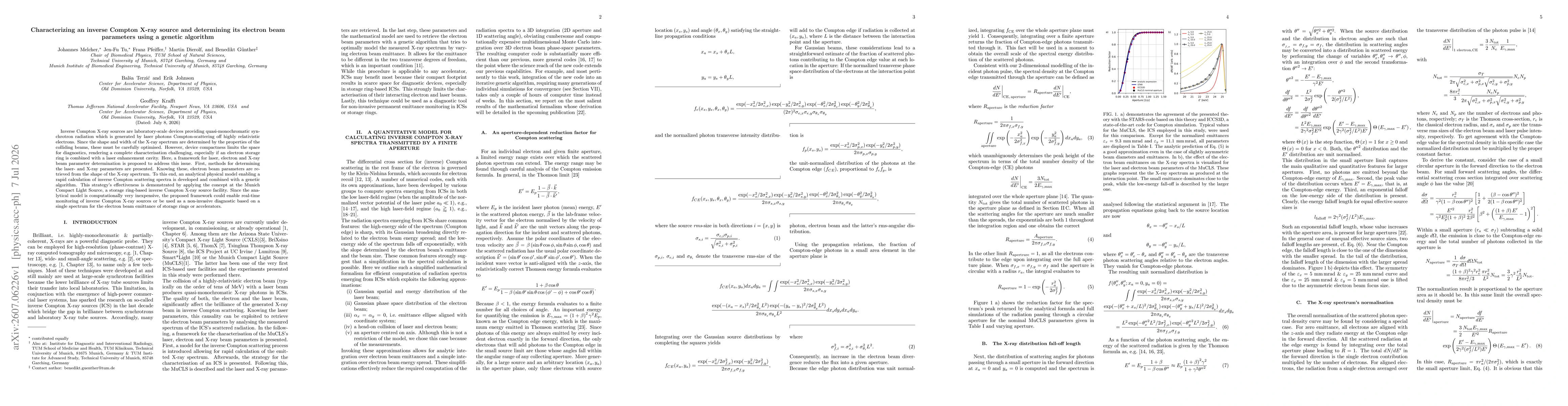
Inverse Compton X-ray sources are laboratory-scale devices providing quasi-monochromatic synchrotron radiation which is generated by laser photons Compton-scattering off highly relativistic electrons....