Technical Note: Low-Dose Simulation for Grating-Based X-Ray Dark-Field Radiography Using a Virtually Decreased Irradiation Area
Publication
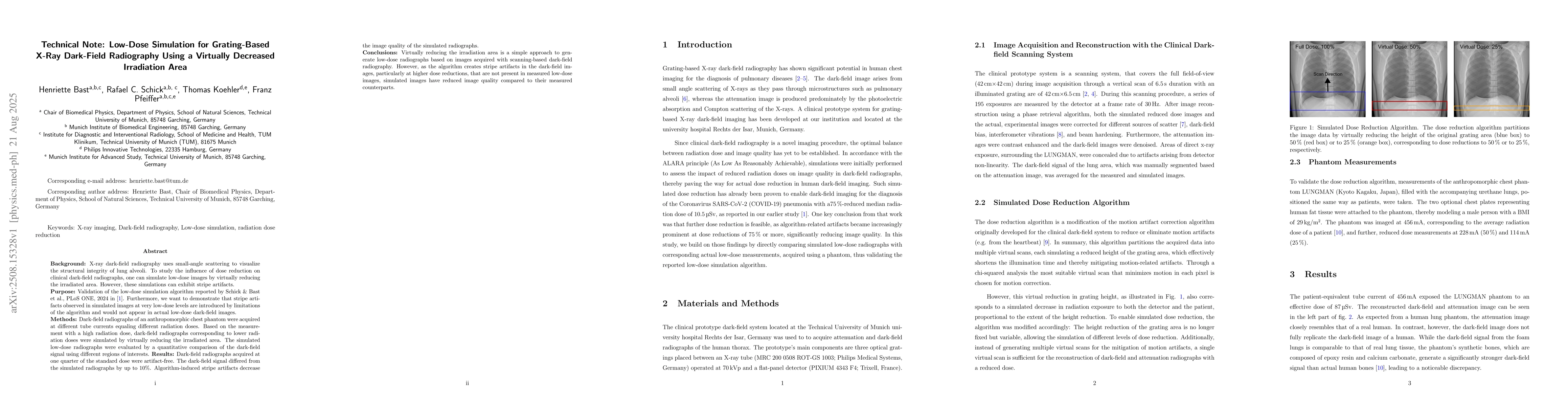
Metrics
Paper Preview
Abstract
Background: X-ray dark-field radiography uses small-angle scattering to visualize the structural integrity of lung alveoli. To study the influence of dose reduction on clinical dark-field radiographs, one can simulate low-dose images by virtually reducing the irradiated area. However, these simulations can exhibit stripe artifacts. Purpose: Validation of the low-dose simulation algorithm reported by Schick & Bast et al., PLoS ONE, 2024. Furthermore, we want to demonstrate that stripe artifacts observed in simulated images at very low-dose levels are introduced by limitations of the algorithm and would not appear in actual low-dose dark-field images. Methods: Dark-field radiographs of an anthropomorphic chest phantom were acquired at different tube currents equaling different radiation doses. Based on the measurement with a high radiation dose, dark-field radiographs corresponding to lower radiation doses were simulated by virtually reducing the irradiated area. The simulated low-dose radiographs were evaluated by a quantitative comparison of the dark-field signal using different regions of interests. Results: Dark-field radiographs acquired at one quarter of the standard dose were artifact-free. The dark-field signal differed from the simulated radiographs by up to 10%. Algorithm-induced stripe artifacts decrease the image quality of the simulated radiographs. Conclusions: Virtually reducing the irradiation area is a simple approach to generate low-dose radiographs based on images acquired with scanning-based dark-field radiography. However, as the algorithm creates stripe artifacts in the dark-field images, particularly at higher dose reductions, that are not present in measured low-dose images, simulated images have reduced image quality compared to their measured counterparts.
AI Key Findings
Get AI-generated insights about this paper's methodology, results, significance, and more — seven facets brought into focus.
Discussion 0