The contribution of T2 relaxation time to diffusion MRI quantification and its clinical implications: a hypothesis
Publication
Metrics
AI Quick Summary
This paper hypothesizes that the longer T2 relaxation time in organs like the spleen and tumors may mitigate signal decay in diffusion MRI, leading to underestimated diffusion values. It suggests that this phenomenon could explain why tumors often show lower apparent diffusion coefficient (ADC) and slow diffusion (Dslow) despite their oedematous nature.
Paper Preview
Abstract
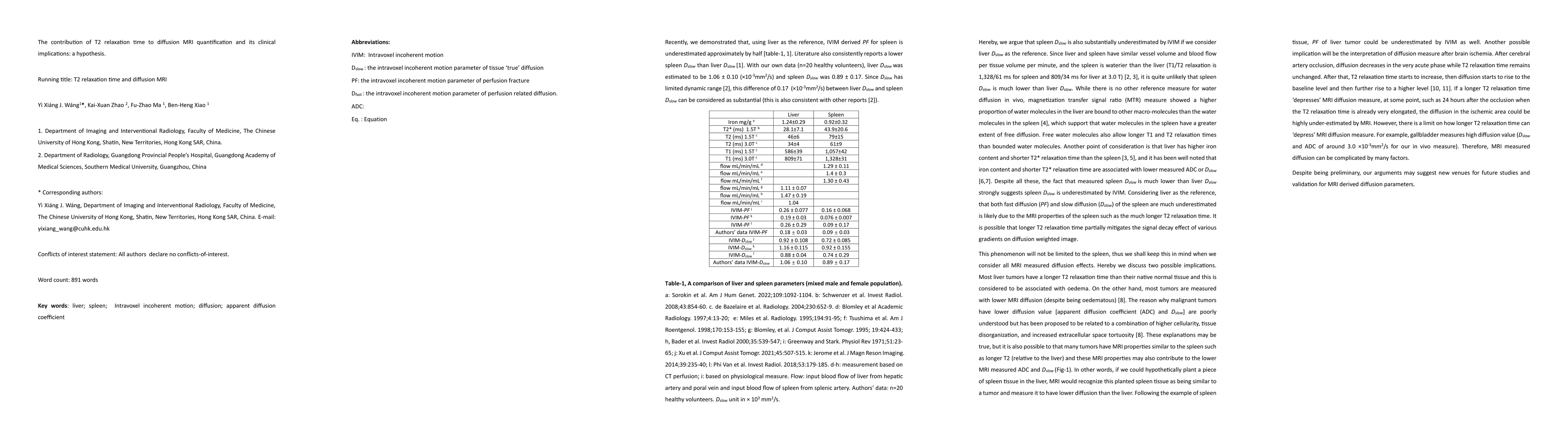
Considering liver as the reference, that both fast diffusion (PF) and slow diffusion (Dslow) of the spleen are much underestimated is likely due to the MRI properties of the spleen such as the much longer T2 relaxation time. It is possible that longer T2 relaxation time partially mitigates the signal decay effect of various gradients on diffusion weighted image. This phenomenon will not be limited to the spleen. Most liver tumors have a longer T2 relaxation time than their native normal tissue and this is considered to be associated with oedema. On the other hand, most tumors are measured with lower MRI diffusion (despite being oedematous). The reason why malignant tumors have lower diffusion value [apparent diffusion coefficient (ADC) and Dslow] are poorly understood but has been proposed to be related to a combination of higher cellularity, tissue disorganization, and increased extracellular space tortuosity. These explanations may be true, but it is also possible to that many tumors have MRI properties similar to the spleen such as longer T2 (relative to the liver) and these MRI properties may also contribute to the lower MRI measured ADC and Dslow . In other words, if we could hypothetically plant a piece of spleen tissue in the liver, MRI would recognize this planted spleen tissue as being similar to a tumor and measure it to have lower diffusion than the liver.
AI Key Findings
Get AI-generated insights about this paper's methodology, results, significance, and more — seven facets brought into focus.
Impact
Paper Details
Authors
PDF Preview
Key Terms
Citation Network
Current paper (gray), citations (green), references (blue)
Display is limited for performance on very large graphs.
Discussion 0